 ムービー
ムービー コントローラー


+ データを開く

- 基本情報
基本情報
| 登録情報 | データベース: EMDB / ID: EMD-7098 | |||||||||
|---|---|---|---|---|---|---|---|---|---|---|
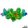
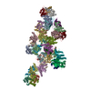
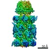
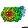
| タイトル | Singly PafE-capped 20S CP in Mycobacterium tuberculosis | |||||||||
 マップデータ マップデータ | Singly PafE-capped 20S CP | |||||||||
 試料 試料 |
| |||||||||
 キーワード キーワード |  Protein degradation (タンパク質分解) / HYDROLASE (加水分解酵素) Protein degradation (タンパク質分解) / HYDROLASE (加水分解酵素) | |||||||||
| 機能・相同性 |  機能・相同性情報 機能・相同性情報symbiont-mediated perturbation of host defenses / proteasome accessory complex / positive regulation of proteasomal protein catabolic process / zymogen binding / proteasome binding / proteasome endopeptidase complex / proteasome core complex, beta-subunit complex / proteasome core complex, alpha-subunit complex / threonine-type endopeptidase activity / regulation of proteasomal protein catabolic process ...symbiont-mediated perturbation of host defenses / proteasome accessory complex / positive regulation of proteasomal protein catabolic process / zymogen binding / proteasome binding / proteasome endopeptidase complex / proteasome core complex, beta-subunit complex / proteasome core complex, alpha-subunit complex / threonine-type endopeptidase activity / regulation of proteasomal protein catabolic process / proteasome complex / proteolysis involved in protein catabolic process / peptidoglycan-based cell wall / proteasomal protein catabolic process / modification-dependent protein catabolic process / protein homooligomerization / extracellular region / 細胞膜 / 細胞質類似検索 - 分子機能 | |||||||||
| 生物種 |   Mycobacterium tuberculosis (結核菌) Mycobacterium tuberculosis (結核菌) | |||||||||
| 手法 | 単粒子再構成法 / クライオ電子顕微鏡法 / 解像度: 4.2 Å | |||||||||
 データ登録者 データ登録者 | Li H / Hu K | |||||||||
 引用 引用 | ジャーナル: J Biol Chem / 年: 2018 タイトル: Proteasome substrate capture and gate opening by the accessory factor PafE from . 著者: Kuan Hu / Jordan B Jastrab / Susan Zhang / Amanda Kovach / Gongpu Zhao / K Heran Darwin / Huilin Li /  要旨: In all domains of life, proteasomes are gated, chambered proteases that require opening by activators to facilitate protein degradation. Twelve proteasome accessory factor E (PafE) monomers assemble ...In all domains of life, proteasomes are gated, chambered proteases that require opening by activators to facilitate protein degradation. Twelve proteasome accessory factor E (PafE) monomers assemble into a single dodecameric ring that promotes proteolysis required for the full virulence of the human bacterial pathogen Whereas the best characterized proteasome activators use ATP to deliver proteins into a proteasome, PafE does not require ATP. Here, to unravel the mechanism of PafE-mediated protein targeting and proteasome activation, we studied the interactions of PafE with native substrates, including a newly identified proteasome substrate, the ParA-like protein, Rv3213c, and with proteasome core particles. We characterized the function of a highly conserved feature in bacterial proteasome activator proteins: a glycine-glutamine-tyrosine-leucine (GQYL) motif at their C termini that is essential for stimulating proteolysis. Using cryo-electron microscopy (cryo-EM), we found that the GQYL motif of PafE interacts with specific residues in the α subunits of the proteasome core particle to trigger gate opening and degradation. Finally, we also found that PafE rings have 40-Å openings lined with hydrophobic residues that form a chamber for capturing substrates before they are degraded, suggesting PafE has a previously unrecognized chaperone activity. In summary, we have identified the interactions between PafE and the proteasome core particle that cause conformational changes leading to the opening of the proteasome gate and have uncovered a mechanism of PafE-mediated substrate degradation. Collectively, our results provide detailed insights into the mechanism of ATP-independent proteasome degradation in bacteria. | |||||||||
| 履歴 |
|
- 構造の表示
構造の表示
| ムービー |
ムービービューア |
|---|---|
| 構造ビューア | EMマップ: SurfViewMolmilJmol/JSmol |
| 添付画像 |

- ダウンロードとリンク
-EMDBアーカイブ
| マップデータ | emd_7098.map.gz | 193.4 MB | EMDBマップデータ形式 | |
|---|---|---|---|---|
| ヘッダ (付随情報) | emd-7098-v30.xmlemd-7098.xml | 11.6 KB 11.6 KB | 表示 表示 | EMDBヘッダ |
| FSC (解像度算出) | emd_7098_fsc.xml | 13.9 KB | 表示 | FSCデータファイル |
| 画像 | 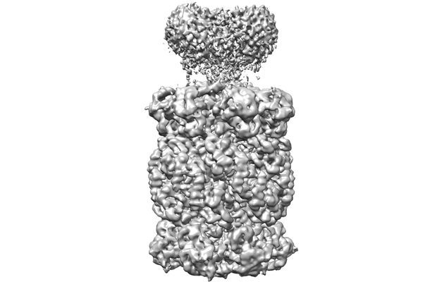 emd_7098.png emd_7098.png | 66.8 KB | ||
| Filedesc metadata | emd-7098.cif.gz | 5.1 KB | ||
| アーカイブディレクトリ |  http://ftp.pdbj.org/pub/emdb/structures/EMD-7098ftp://ftp.pdbj.org/pub/emdb/structures/EMD-7098 http://ftp.pdbj.org/pub/emdb/structures/EMD-7098ftp://ftp.pdbj.org/pub/emdb/structures/EMD-7098 | HTTPS FTP |
-関連構造データ

-リンク
| EMDBのページ | EMDB (EBI/PDBe) / EMDataResource |
|---|---|
| 「今月の分子」の関連する項目 |


-マップ
| ファイル | ダウンロード / ファイル: emd_7098.map.gz / 形式: CCP4 / 大きさ: 244.1 MB / タイプ: IMAGE STORED AS FLOATING POINT NUMBER (4 BYTES) | ||||||||||||||||||||||||||||||||||||||||||||||||||||||||||||
|---|---|---|---|---|---|---|---|---|---|---|---|---|---|---|---|---|---|---|---|---|---|---|---|---|---|---|---|---|---|---|---|---|---|---|---|---|---|---|---|---|---|---|---|---|---|---|---|---|---|---|---|---|---|---|---|---|---|---|---|---|---|
| 注釈 | Singly PafE-capped 20S CP | ||||||||||||||||||||||||||||||||||||||||||||||||||||||||||||
| ボクセルのサイズ | X=Y=Z: 1.09 Å | ||||||||||||||||||||||||||||||||||||||||||||||||||||||||||||
| 密度 |
| ||||||||||||||||||||||||||||||||||||||||||||||||||||||||||||
| 対称性 | 空間群: 1 | ||||||||||||||||||||||||||||||||||||||||||||||||||||||||||||
| 詳細 | EMDB XML:
CCP4マップ ヘッダ情報:
| ||||||||||||||||||||||||||||||||||||||||||||||||||||||||||||
-添付データ
- 試料の構成要素
-全体 : Singly PafE-capped 20S CP
| 全体 | 名称: Singly PafE-capped 20S CP |
|---|---|
| 要素 |
|
-超分子 #1: Singly PafE-capped 20S CP
| 超分子 | 名称: Singly PafE-capped 20S CP / タイプ: complex / ID: 1 / 親要素: 0 / 含まれる分子: all |
|---|---|
| 由来(天然) | 生物種: Mycobacterium tuberculosis (結核菌) |
-分子 #1: Proteasome subunit alpha
| 分子 | 名称: Proteasome subunit alpha / タイプ: protein_or_peptide / ID: 1 / コピー数: 14 / 光学異性体: LEVO / EC番号: proteasome endopeptidase complex |
|---|---|
| 由来(天然) | 生物種: Mycobacterium tuberculosis (結核菌) |
| 分子量 | 理論値: 26.911039 KDa |
| 組換発現 | 生物種: Escherichia coli (大腸菌) |
| 配列 | 文字列: MSFPYFISPE QAMRERSELA RKGIARAKSV VALAYAGGVL FVAENPSRSL QKISELYDRV GFAAAGKFNE FDNLRRGGIQ FADTRGYAY DRRDVTGRQL ANVYAQTLGT IFTEQAKPYE VELCVAEVAH YGETKRPELY RITYDGSIAD EPHFVVMGGT T EPIANALK ...文字列: MSFPYFISPE QAMRERSELA RKGIARAKSV VALAYAGGVL FVAENPSRSL QKISELYDRV GFAAAGKFNE FDNLRRGGIQ FADTRGYAY DRRDVTGRQL ANVYAQTLGT IFTEQAKPYE VELCVAEVAH YGETKRPELY RITYDGSIAD EPHFVVMGGT T EPIANALK ESYAENASLT DALRIAVAAL RAGSADTSGG DQPTLGVASL EVAVLDANRP RRAFRRITGS ALQALLVDQE SP QSDGESS G UniProtKB: Proteasome subunit alpha |
-分子 #2: Proteasome subunit beta
| 分子 | 名称: Proteasome subunit beta / タイプ: protein_or_peptide / ID: 2 / コピー数: 14 / 光学異性体: LEVO / EC番号: proteasome endopeptidase complex |
|---|---|
| 由来(天然) | 生物種: Mycobacterium tuberculosis (結核菌) |
| 分子量 | 理論値: 25.274264 KDa |
| 組換発現 | 生物種: Escherichia coli (大腸菌) |
| 配列 | 文字列: TTIVALKYPG GVVMAGDRRS TQGNMISGRD VRKVYITDDY TATGIAGTAA VAVEFARLYA VELEHYEKLE GVPLTFAGKI NRLAIMVRG NLAAAMQGLL ALPLLAGYDI HASDPQSAGR IVSFDAAGGW NIEEEGYQAV GSGSLFAKSS MKKLYSQVTD G DSGLRVAV ...文字列: TTIVALKYPG GVVMAGDRRS TQGNMISGRD VRKVYITDDY TATGIAGTAA VAVEFARLYA VELEHYEKLE GVPLTFAGKI NRLAIMVRG NLAAAMQGLL ALPLLAGYDI HASDPQSAGR IVSFDAAGGW NIEEEGYQAV GSGSLFAKSS MKKLYSQVTD G DSGLRVAV EALYDAADDD SATGGPDLVR GIFPTAVIID ADGAVDVPES RIAELARAII ESRSGADTFG SDGGEKHHHH HH UniProtKB: Proteasome subunit beta |
-分子 #3: Bacterial proteasome activator
| 分子 | 名称: Bacterial proteasome activator / タイプ: protein_or_peptide / ID: 3 / コピー数: 7 / 光学異性体: LEVO |
|---|---|
| 由来(天然) | 生物種: Mycobacterium tuberculosis (結核菌) |
| 分子量 | 理論値: 18.963232 KDa |
| 組換発現 | 生物種: Escherichia coli (大腸菌) |
| 配列 | 文字列: MVIGLSTGSD DDDVEVIGGV DPRLIAVQEN DSDESSLTDL VEQPAKVMRI GTMIKQLLEE VRAAPLDEAS RNRLRDIHAT SIRELEDGL APELREELDR LTLPFNEDAV PSDAELRIAQ AQLVGWLEGL FHGIQTALFA QQMAARAQLQ QMRQGALPPG V GKSGQHGH GTGQYL UniProtKB: Bacterial proteasome activator |
-実験情報
-構造解析
| 手法 | クライオ電子顕微鏡法 |
|---|---|
 解析 解析 | 単粒子再構成法 |
| 試料の集合状態 | particle |
-試料調製
| 緩衝液 | pH: 7.4 |
|---|---|
| 凍結 | 凍結剤: ETHANE |
- 電子顕微鏡法
| 顕微鏡 | FEI TITAN KRIOS |
|---|---|
| 電子線 | 加速電圧: 300 kV / 電子線源: FIELD EMISSION GUN |
| 電子光学系 | 照射モード: FLOOD BEAM / 撮影モード: BRIGHT FIELDBright-field microscopy |
| 撮影 | フィルム・検出器のモデル: GATAN K2 SUMMIT (4k x 4k) 平均電子線量: 1.9 e/Å2 |
| 実験機器 |  モデル: Titan Krios / 画像提供: FEI Company |
-画像解析
| 初期モデル | モデルのタイプ: PDB ENTRY |
|---|---|
| 初期 角度割当 | タイプ: ANGULAR RECONSTITUTION |
| 最終 角度割当 | タイプ: ANGULAR RECONSTITUTION |
| 最終 再構成 | 想定した対称性 - 点群: C1 (非対称) / 解像度のタイプ: BY AUTHOR / 解像度: 4.2 Å / 解像度の算出法: FSC 0.143 CUT-OFF / 使用した粒子像数: 60034 |
| FSC曲線 (解像度の算出) | 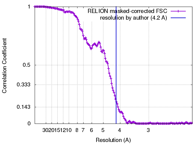 |
