 Movie
Movie Controller

[English] 日本語
 Yorodumi
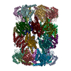
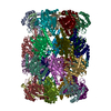
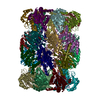
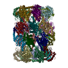
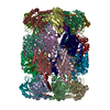
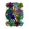
- PDB-6bgl: Doubly PafE-capped 20S core particle in Mycobacterium tuberculosis -
+ Open data

- Basic information
| Entry | Database: PDB / ID: 6bgl | ||||||
|---|---|---|---|---|---|---|---|
| Title | Doubly PafE-capped 20S core particle in Mycobacterium tuberculosis | ||||||
 Components Components |
| ||||||
 Keywords Keywords | HYDROLASE / Protein degradation | ||||||
| Function / homology |  Function and homology information Function and homology informationsymbiont-mediated perturbation of host defenses / zymogen binding / proteasome accessory complex / positive regulation of proteasomal protein catabolic process / proteasome binding / proteasome endopeptidase complex / proteasome core complex, beta-subunit complex / threonine-type endopeptidase activity / proteasome core complex, alpha-subunit complex / peptidoglycan-based cell wall ...symbiont-mediated perturbation of host defenses / zymogen binding / proteasome accessory complex / positive regulation of proteasomal protein catabolic process / proteasome binding / proteasome endopeptidase complex / proteasome core complex, beta-subunit complex / threonine-type endopeptidase activity / proteasome core complex, alpha-subunit complex / peptidoglycan-based cell wall / regulation of proteasomal protein catabolic process / : / proteasome complex / proteasomal protein catabolic process / protein homooligomerization / modification-dependent protein catabolic process / proteasome-mediated ubiquitin-dependent protein catabolic process / extracellular region / plasma membrane / cytoplasm Similarity search - Function | ||||||
| Biological species |   Mycobacterium tuberculosis (bacteria) Mycobacterium tuberculosis (bacteria) | ||||||
| Method | ELECTRON MICROSCOPY / single particle reconstruction / cryo EM / Resolution: 3.4 Å | ||||||
 Authors Authors | Li, H. / Hu, K. | ||||||
 Citation Citation | Journal: J Biol Chem / Year: 2018 Title: Proteasome substrate capture and gate opening by the accessory factor PafE from . Authors: Kuan Hu / Jordan B Jastrab / Susan Zhang / Amanda Kovach / Gongpu Zhao / K Heran Darwin / Huilin Li /  Abstract: In all domains of life, proteasomes are gated, chambered proteases that require opening by activators to facilitate protein degradation. Twelve proteasome accessory factor E (PafE) monomers assemble ...In all domains of life, proteasomes are gated, chambered proteases that require opening by activators to facilitate protein degradation. Twelve proteasome accessory factor E (PafE) monomers assemble into a single dodecameric ring that promotes proteolysis required for the full virulence of the human bacterial pathogen Whereas the best characterized proteasome activators use ATP to deliver proteins into a proteasome, PafE does not require ATP. Here, to unravel the mechanism of PafE-mediated protein targeting and proteasome activation, we studied the interactions of PafE with native substrates, including a newly identified proteasome substrate, the ParA-like protein, Rv3213c, and with proteasome core particles. We characterized the function of a highly conserved feature in bacterial proteasome activator proteins: a glycine-glutamine-tyrosine-leucine (GQYL) motif at their C termini that is essential for stimulating proteolysis. Using cryo-electron microscopy (cryo-EM), we found that the GQYL motif of PafE interacts with specific residues in the α subunits of the proteasome core particle to trigger gate opening and degradation. Finally, we also found that PafE rings have 40-Å openings lined with hydrophobic residues that form a chamber for capturing substrates before they are degraded, suggesting PafE has a previously unrecognized chaperone activity. In summary, we have identified the interactions between PafE and the proteasome core particle that cause conformational changes leading to the opening of the proteasome gate and have uncovered a mechanism of PafE-mediated substrate degradation. Collectively, our results provide detailed insights into the mechanism of ATP-independent proteasome degradation in bacteria. | ||||||
| History |
|
- Structure visualization
| Movie |
Movie viewer |
|---|---|
| Structure viewer | Molecule: MolmilJmol/JSmol |
- Downloads & links
-Download
| PDBx/mmCIF format | 6bgl.cif.gz | 1.1 MB | Display | PDBx/mmCIF format |
|---|---|---|---|---|
| PDB format | pdb6bgl.ent.gz | 942.6 KB | Display | PDB format |
| PDBx/mmJSON format | 6bgl.json.gz | Tree view | PDBx/mmJSON format | |
| Others |  Other downloads Other downloads |
-Validation report
| Arichive directory | https://data.pdbj.org/pub/pdb/validation_reports/bg/6bglftp://data.pdbj.org/pub/pdb/validation_reports/bg/6bgl | HTTPS FTP |
|---|
-Related structure data
| Related structure data | 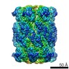 7097MC  7098C 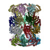 6bgoC M: map data used to model this data C: citing same article ( |
|---|---|
| Similar structure data |


-Links
 PDBj
PDBj


- Assembly
| Deposited unit | 
|
|---|---|
| 1 |
|
-Components
| #1: Protein | Mass: 26911.039 Da / Num. of mol.: 14 Source method: isolated from a genetically manipulated source Source: (gene. exp.) Mycobacterium tuberculosis (bacteria) / Gene: prcA, MRA_2124 / Production host: References: UniProt: A5U4D5, UniProt: P9WHU1*PLUS, proteasome endopeptidase complex #2: Protein | Mass: 18963.232 Da / Num. of mol.: 14 Source method: isolated from a genetically manipulated source Source: (gene. exp.) Mycobacterium tuberculosis (bacteria) / Gene: bpa, MT3889 / Production host: #3: Protein | Mass: 25274.264 Da / Num. of mol.: 14 Source method: isolated from a genetically manipulated source Source: (gene. exp.) Mycobacterium tuberculosis (bacteria) / Gene: prcB, MRA_2125 / Production host: References: UniProt: A5U4D6, UniProt: P9WHT9*PLUS, proteasome endopeptidase complex |
|---|
-Experimental details
-Experiment
| Experiment | Method: ELECTRON MICROSCOPY |
|---|---|
| EM experiment | Aggregation state: PARTICLE / 3D reconstruction method: single particle reconstruction |
- Sample preparation
| Component | Name: Doubly PafE-capped 20S CP / Type: COMPLEX / Entity ID: all / Source: RECOMBINANT |
|---|---|
| Source (natural) | Organism: Mycobacterium tuberculosis (bacteria) |
| Source (recombinant) | Organism: |
| Buffer solution | pH: 7.4 |
| Specimen | Embedding applied: NO / Shadowing applied: NO / Staining applied: NO / Vitrification applied: YES |
| Vitrification | Cryogen name: ETHANE |
- Electron microscopy imaging
| Experimental equipment |  Model: Titan Krios / Image courtesy: FEI Company |
|---|---|
| Microscopy | Model: FEI TITAN KRIOS |
| Electron gun | Electron source:  FIELD EMISSION GUN / Accelerating voltage: 300 kV / Illumination mode: FLOOD BEAM FIELD EMISSION GUN / Accelerating voltage: 300 kV / Illumination mode: FLOOD BEAM |
| Electron lens | Mode: BRIGHT FIELD |
| Image recording | Electron dose: 1.9 e/Å2 / Film or detector model: GATAN K2 SUMMIT (4k x 4k) |
- Processing
| CTF correction | Type: PHASE FLIPPING AND AMPLITUDE CORRECTION |
|---|---|
| Symmetry | Point symmetry: D7 (2x7 fold dihedral) |
| 3D reconstruction | Resolution: 3.4 Å / Resolution method: FSC 0.143 CUT-OFF / Num. of particles: 51091 / Symmetry type: POINT |
