ムービー
ムービー コントローラー


+ データを開く

- 基本情報
基本情報
| 登録情報 | データベース: EMDB / ID: EMD-6385 | |||||||||
|---|---|---|---|---|---|---|---|---|---|---|
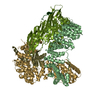
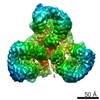
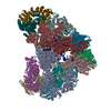
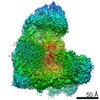
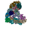
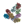
| タイトル | Electron cryo-microscopy of AP-1:Arf1:Nef complex | |||||||||
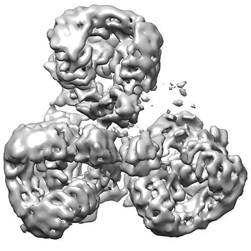
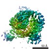
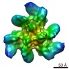
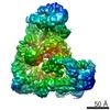
 マップデータ マップデータ | Trimeric structure of AP-1:Arf1:Nef | |||||||||
 試料 試料 |
| |||||||||
 キーワード キーワード |  adaptor protein-1 / Arf1 / Nef / clathrin cage adaptor protein-1 / Arf1 / Nef / clathrin cage | |||||||||
| 生物種 |  Homo sapiens (ヒト) Homo sapiens (ヒト) | |||||||||
| 手法 | 単粒子再構成法 / クライオ電子顕微鏡法 / 解像度: 7.0 Å | |||||||||
 データ登録者 データ登録者 | Shen QT / Ren X / Zhang R / Lee H / Hurley J | |||||||||
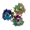
 引用 引用 | ジャーナル: Science / 年: 2015 タイトル: HIV-1 Nef hijacks clathrin coats by stabilizing AP-1:Arf1 polygons. 著者: Qing-Tao Shen / Xuefeng Ren / Rui Zhang / Il-Hyung Lee / James H Hurley /  要旨: The lentiviruses HIV and simian immunodeficiency virus (SIV) subvert intracellular membrane traffic as part of their replication cycle. The lentiviral Nef protein helps viruses evade innate and ...The lentiviruses HIV and simian immunodeficiency virus (SIV) subvert intracellular membrane traffic as part of their replication cycle. The lentiviral Nef protein helps viruses evade innate and adaptive immune defenses by hijacking the adaptor protein 1 (AP-1) and AP-2 clathrin adaptors. We found that HIV-1 Nef and the guanosine triphosphatase Arf1 induced trimerization and activation of AP-1. Here we report the cryo-electron microscopy structures of the Nef- and Arf1-bound AP-1 trimer in the active and inactive states. A central nucleus of three Arf1 molecules organizes the trimers. We combined the open trimer with a known dimer structure and thus predicted a hexagonal assembly with inner and outer faces that bind the membranes and clathrin, respectively. Hexagons were directly visualized and the model validated by reconstituting clathrin cage assembly. Arf1 and Nef thus play interconnected roles in allosteric activation, cargo recruitment, and coat assembly, revealing an unexpectedly intricate organization of the inner AP-1 layer of the clathrin coat. | |||||||||
| 履歴 |
|
- 構造の表示
構造の表示
| ムービー |
 ムービービューア ムービービューア |
|---|---|
| 構造ビューア | EMマップ: SurfViewMolmilJmol/JSmol |
| 添付画像 |
- ダウンロードとリンク
-EMDBアーカイブ
| マップデータ | emd_6385.map.gz | 7.4 MB | EMDBマップデータ形式 | |
|---|---|---|---|---|
| ヘッダ (付随情報) | emd-6385-v30.xmlemd-6385.xml | 11.4 KB 11.4 KB | 表示 表示 | EMDBヘッダ |
| 画像 |  emd_6385.jpg emd_6385.jpg | 149.1 KB | ||
| アーカイブディレクトリ |  http://ftp.pdbj.org/pub/emdb/structures/EMD-6385ftp://ftp.pdbj.org/pub/emdb/structures/EMD-6385 http://ftp.pdbj.org/pub/emdb/structures/EMD-6385ftp://ftp.pdbj.org/pub/emdb/structures/EMD-6385 | HTTPS FTP |
-関連構造データ

-リンク
| EMDBのページ | EMDB (EBI/PDBe) / EMDataResource |
|---|
-マップ
| ファイル | ダウンロード / ファイル: emd_6385.map.gz / 形式: CCP4 / 大きさ: 7.8 MB / タイプ: IMAGE STORED AS FLOATING POINT NUMBER (4 BYTES) | ||||||||||||||||||||||||||||||||||||||||||||||||||||||||||||
|---|---|---|---|---|---|---|---|---|---|---|---|---|---|---|---|---|---|---|---|---|---|---|---|---|---|---|---|---|---|---|---|---|---|---|---|---|---|---|---|---|---|---|---|---|---|---|---|---|---|---|---|---|---|---|---|---|---|---|---|---|---|
| 注釈 | Trimeric structure of AP-1:Arf1:Nef | ||||||||||||||||||||||||||||||||||||||||||||||||||||||||||||
| ボクセルのサイズ | X=Y=Z: 2.64 Å | ||||||||||||||||||||||||||||||||||||||||||||||||||||||||||||
| 密度 |
| ||||||||||||||||||||||||||||||||||||||||||||||||||||||||||||
| 対称性 | 空間群: 1 | ||||||||||||||||||||||||||||||||||||||||||||||||||||||||||||
| 詳細 | EMDB XML:
CCP4マップ ヘッダ情報:
| ||||||||||||||||||||||||||||||||||||||||||||||||||||||||||||
-添付データ
- 試料の構成要素
-全体 : AP-1:Arf1:Nef complex
| 全体 | 名称: AP-1:Arf1:Nef complex |
|---|---|
| 要素 |
|
-超分子 #1000: AP-1:Arf1:Nef complex
| 超分子 | 名称: AP-1:Arf1:Nef complex / タイプ: sample / ID: 1000 / 集合状態: trimer / Number unique components: 3 |
|---|---|
| 分子量 | 実験値: 850 KDa / 理論値: 870 KDa / 手法: multi-angle light scattering |
-分子 #1: adaptor protein-1
| 分子 | 名称: adaptor protein-1 / タイプ: protein_or_peptide / ID: 1 / 組換発現: Yes |
|---|---|
| 由来(天然) | 生物種: Homo sapiens (ヒト) / 別称: Human / 細胞中の位置: trans-Golgi network |
| 組換発現 | 生物種:  Escherichia coli (大腸菌) / 組換株: BL21(DE3) pLys / 組換プラスミド: pST44 Escherichia coli (大腸菌) / 組換株: BL21(DE3) pLys / 組換プラスミド: pST44 |
-分子 #2: Arf1
| 分子 | 名称: Arf1 / タイプ: protein_or_peptide / ID: 2 / Name.synonym: ADP-ribosylation factor-1 / 組換発現: Yes |
|---|---|
| 由来(天然) | 生物種: Homo sapiens (ヒト) / 別称: human |
| 分子量 | 実験値: 200 KDa / 理論値: 200 KDa |
| 組換発現 | 生物種: Escherichia coli (大腸菌) / 組換株: BL21(DE3) pLys / 組換プラスミド: pST44 |
-分子 #3: Nef
| 分子 | 名称: Nef / タイプ: protein_or_peptide / ID: 3 / Name.synonym: negative factor / 組換発現: Yes |
|---|---|
| 由来(天然) | 生物種: Homo sapiens (ヒト) / 別称: human |
| 分子量 | 実験値: 290 KDa / 理論値: 290 KDa |
| 組換発現 | 生物種: Escherichia coli (大腸菌) / 組換株: BL21(DE3) pLys / 組換プラスミド: pST44 |
-実験情報
-構造解析
| 手法 | クライオ電子顕微鏡法 |
|---|---|
 解析 解析 | 単粒子再構成法 |
| 試料の集合状態 | particle |
-試料調製
| 濃度 | 0.2 mg/mL |
|---|---|
| 緩衝液 | pH: 8 / 詳細: 20 mM Tris, 200 mM NaCl, 0.3 mM TCEP |
| グリッド | 詳細: 400 mesh copper grid with thin carbon support |
| 凍結 | 凍結剤: ETHANE / チャンバー内湿度: 100 % / チャンバー内温度: 120 K / 装置: FEI VITROBOT MARK IV / 手法: Blot for 2 seconds before plunging |
- 電子顕微鏡法
| 顕微鏡 | FEI TITAN |
|---|---|
| 電子線 | 加速電圧: 300 kV / 電子線源: FIELD EMISSION GUN |
| 電子光学系 | 倍率(補正後): 80000 / 照射モード: SPOT SCAN / 撮影モード: BRIGHT FIELDBright-field microscopy / Cs: 2.7 mm / 最大 デフォーカス(公称値): 2.0 µm / 最小 デフォーカス(公称値): 0.8 µm / 倍率(公称値): 80000 |
| 試料ステージ | 試料ホルダーモデル: GATAN LIQUID NITROGEN |
| 日付 | 2015年3月30日 |
| 撮影 | カテゴリ: CCD フィルム・検出器のモデル: DIRECT ELECTRON DE-12 (4k x 3k) 実像数: 12000 / 平均電子線量: 45.9 e/Å2 |
-画像解析
| 最終 2次元分類 | クラス数: 10 |
|---|---|
| 最終 再構成 | 解像度のタイプ: BY AUTHOR / 解像度: 7.0 Å / 解像度の算出法: OTHER / ソフトウェア - 名称: RELION / 使用した粒子像数: 12000 |
| 詳細 | The particles were picked using the DoG Picker. |
-原子モデル構築 1
| 初期モデル | PDB ID: |
|---|---|
| ソフトウェア | 名称: Chimera |
| 精密化 | 空間: REAL / プロトコル: RIGID BODY FIT |