- EMDB-5201: The electron cryo-microscopic structure of Shigella phage Sf6 rev... -
+
データを開く
IDまたはキーワード:
読み込み中...
-
基本情報
登録情報
データベース: EMDB / ID: EMD-5201
タイトル
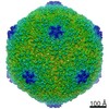
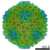
The electron cryo-microscopic structure of Shigella phage Sf6 reveals novel cementing proteins
マップデータ
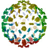
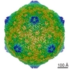
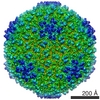
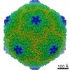
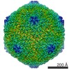
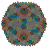
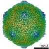
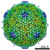
Electron cryo-microscopic reconstruction of Shigella phage Sf6. The image is viewed approximately down the icosahedral 3-fold axis, and an octant of the capsid was removed to show the internal density (colored in green) that corresponds to the two host-derived cementing proteins.
outer membrane protein complex / monoatomic ion transmembrane transporter activity / detection of virus / outer membrane / porin activity / pore complex / monoatomic ion transport / monoatomic ion transmembrane transport / cell outer membrane / virus receptor activity ...outer membrane protein complex / monoatomic ion transmembrane transporter activity / detection of virus / outer membrane / porin activity / pore complex / monoatomic ion transport / monoatomic ion transmembrane transport / cell outer membrane / virus receptor activity / outer membrane-bounded periplasmic space / receptor-mediated virion attachment to host cell / symbiont entry into host cell / DNA damage response / 生体膜 / identical protein binding / metal ion binding 類似検索 - 分子機能
Outer membrane protein OmpA-like, transmembrane domain / Outer membrane protein, OmpA / OmpA-like transmembrane domain / Outer membrane protein, OmpA-like, conserved site / OmpA-like domain. / Outer membrane protein, bacterial / Porin, gammaproteobacterial / Porin, Gram-negative type, conserved site / General diffusion Gram-negative porins signature. / Gram-negative porin ...Outer membrane protein OmpA-like, transmembrane domain / Outer membrane protein, OmpA / OmpA-like transmembrane domain / Outer membrane protein, OmpA-like, conserved site / OmpA-like domain. / Outer membrane protein, bacterial / Porin, gammaproteobacterial / Porin, Gram-negative type, conserved site / General diffusion Gram-negative porins signature. / Gram-negative porin / Porin, Gram-negative type / OmpA-like domain superfamily / OmpA family / OmpA-like domain / OmpA-like domain profile. / Porin domain superfamily / Outer membrane protein/outer membrane enzyme PagP, beta-barrel 類似検索 - ドメイン・相同性
Outer membrane porin C / Outer membrane protein A 類似検索 - 構成要素
ジャーナル: Virology / 年: 2011 タイトル: The host outer membrane proteins OmpA and OmpC are associated with the Shigella phage Sf6 virion. 著者: Haiyan Zhao / Reuben D Sequeira / Nadezhda A Galeva / Liang Tang / 要旨: Assembly of dsDNA bacteriophage is a precisely programmed process. Potential roles of host cell components in phage assembly haven't been well understood. It was previously reported that two ...Assembly of dsDNA bacteriophage is a precisely programmed process. Potential roles of host cell components in phage assembly haven't been well understood. It was previously reported that two unidentified proteins were present in bacteriophage Sf6 virion (Casjens et al, 2004, J.Mol.Biol. 339, 379-394, Fig. 2A). Using tandem mass spectrometry, we have identified the two proteins as outer membrane proteins (OMPs) OmpA and OmpC from its host Shigella flexneri. The transmission electron cryo-microscopy structure of Sf6 shows significant density at specific sites at the phage capsid inner surface. This density fit well with the characteristic beta-barrel domains of OMPs, thus may be due to the two host proteins. Locations of this density suggest a role in Sf6 morphogenesis reminiscent of phage-encoded cementing proteins. These data indicate a new, OMP-related phage:host linkage, adding to previous knowledge that some lambdoid bacteriophage genomes contain OmpC-like genes that express phage-encoded porins in the lysogenic state.
ダウンロード / ファイル: emd_5201.map.gz / 形式: CCP4 / 大きさ: 29.8 MB / タイプ: IMAGE STORED AS FLOATING POINT NUMBER (4 BYTES)
注釈
Electron cryo-microscopic reconstruction of Shigella phage Sf6. The image is viewed approximately down the icosahedral 3-fold axis, and an octant of the capsid was removed to show the internal density (colored in green) that corresponds to the two host-derived cementing proteins.
 ムービー
ムービー コントローラー


 データを開く
 基本情報
基本情報 マップデータ
 試料
試料 キーワード
キーワード virus assembly (ウイルス) / cementing protein /
  機能・相同性情報
 Bacillus phage SF6 (ファージ)
 データ登録者
 引用
引用
 構造の表示
構造の表示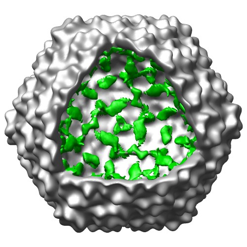
 ダウンロードとリンク
 emd_5201_1.jpg
 http://ftp.pdbj.org/pub/emdb/structures/EMD-5201
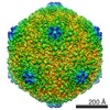


 試料の構成要素
 解析
解析 電子顕微鏡法