 ムービー
ムービー コントローラー


+ データを開く

- 基本情報
基本情報
| 登録情報 | データベース: EMDB / ID: EMD-3559 | |||||||||
|---|---|---|---|---|---|---|---|---|---|---|
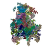
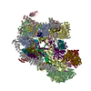
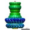
| タイトル | mitochondrial ATP synthase dimer from Euglena gracilis | |||||||||
 マップデータ マップデータ | subtomogram average of mitochondrial ATP synthase dimer from Euglena gracilis | |||||||||
 試料 試料 |
| |||||||||
| 生物種 |   Euglena gracilis (ミドリムシ) Euglena gracilis (ミドリムシ) | |||||||||
| 手法 | サブトモグラム平均法 / クライオ電子顕微鏡法 / 解像度: 27.5 Å | |||||||||
 データ登録者 データ登録者 | Muehleip AW / Davies KM / Kuehlbrandt W | |||||||||
 引用 引用 | ジャーナル: Proc Natl Acad Sci U S A / 年: 2017 タイトル: In situ structure of trypanosomal ATP synthase dimer reveals a unique arrangement of catalytic subunits. 著者: Alexander W Mühleip / Caroline E Dewar / Achim Schnaufer / Werner Kühlbrandt / Karen M Davies /   要旨: We used electron cryotomography and subtomogram averaging to determine the in situ structures of mitochondrial ATP synthase dimers from two organisms belonging to the phylum euglenozoa: Trypanosoma ...We used electron cryotomography and subtomogram averaging to determine the in situ structures of mitochondrial ATP synthase dimers from two organisms belonging to the phylum euglenozoa: Trypanosoma brucei, a lethal human parasite, and Euglena gracilis, a photosynthetic protist. At a resolution of 32.5 Å and 27.5 Å, respectively, the two structures clearly exhibit a noncanonical F head, in which the catalytic (αβ) assembly forms a triangular pyramid rather than the pseudo-sixfold ring arrangement typical of all other ATP synthases investigated so far. Fitting of known X-ray structures reveals that this unusual geometry results from a phylum-specific cleavage of the α subunit, in which the C-terminal α fragments are displaced by ∼20 Å and rotated by ∼30° from their expected positions. In this location, the α fragment is unable to form the conserved catalytic interface that was thought to be essential for ATP synthesis, and cannot convert γ-subunit rotation into the conformational changes implicit in rotary catalysis. The new arrangement of catalytic subunits suggests that the mechanism of ATP generation by rotary ATPases is less strictly conserved than has been generally assumed. The ATP synthases of these organisms present a unique model system for discerning the individual contributions of the α and β subunits to the fundamental process of ATP synthesis. | |||||||||
| 履歴 |
|
- 構造の表示
構造の表示
| ムービー |
 ムービービューア ムービービューア |
|---|---|
| 構造ビューア | EMマップ: SurfViewMolmilJmol/JSmol |
| 添付画像 |

- ダウンロードとリンク
-EMDBアーカイブ
| マップデータ | emd_3559.map.gz | 22.1 MB | EMDBマップデータ形式 | |
|---|---|---|---|---|
| ヘッダ (付随情報) | emd-3559-v30.xmlemd-3559.xml | 11.1 KB 11.1 KB | 表示 表示 | EMDBヘッダ |
| 画像 |  emd_3559.png emd_3559.png | 83.4 KB | ||
| アーカイブディレクトリ |  http://ftp.pdbj.org/pub/emdb/structures/EMD-3559ftp://ftp.pdbj.org/pub/emdb/structures/EMD-3559 http://ftp.pdbj.org/pub/emdb/structures/EMD-3559ftp://ftp.pdbj.org/pub/emdb/structures/EMD-3559 | HTTPS FTP |
-関連構造データ

-リンク
| EMDBのページ | EMDB (EBI/PDBe) / EMDataResource |
|---|
-マップ
| ファイル | ダウンロード / ファイル: emd_3559.map.gz / 形式: CCP4 / 大きさ: 30.5 MB / タイプ: IMAGE STORED AS FLOATING POINT NUMBER (4 BYTES) | ||||||||||||||||||||||||||||||||||||||||||||||||||||||||||||
|---|---|---|---|---|---|---|---|---|---|---|---|---|---|---|---|---|---|---|---|---|---|---|---|---|---|---|---|---|---|---|---|---|---|---|---|---|---|---|---|---|---|---|---|---|---|---|---|---|---|---|---|---|---|---|---|---|---|---|---|---|---|
| 注釈 | subtomogram average of mitochondrial ATP synthase dimer from Euglena gracilis | ||||||||||||||||||||||||||||||||||||||||||||||||||||||||||||
| ボクセルのサイズ | X=Y=Z: 4.56 Å | ||||||||||||||||||||||||||||||||||||||||||||||||||||||||||||
| 密度 |
| ||||||||||||||||||||||||||||||||||||||||||||||||||||||||||||
| 対称性 | 空間群: 1 | ||||||||||||||||||||||||||||||||||||||||||||||||||||||||||||
| 詳細 | EMDB XML:
CCP4マップ ヘッダ情報:
| ||||||||||||||||||||||||||||||||||||||||||||||||||||||||||||
-添付データ
- 試料の構成要素
-全体 : ATP synthase dimer from Euglena gracilis in isolated mitochondria...
| 全体 | 名称: ATP synthase dimer from Euglena gracilis in isolated mitochondrial membranes |
|---|---|
| 要素 |
|
-超分子 #1: ATP synthase dimer from Euglena gracilis in isolated mitochondria...
| 超分子 | 名称: ATP synthase dimer from Euglena gracilis in isolated mitochondrial membranes タイプ: organelle_or_cellular_component / ID: 1 / 親要素: 0 |
|---|---|
| 由来(天然) | 生物種: Euglena gracilis (ミドリムシ) / Organelle: mitochondrion |
| 分子量 | 理論値: 2 MDa |
-実験情報
-構造解析
| 手法 | クライオ電子顕微鏡法 |
|---|---|
 解析 解析 | サブトモグラム平均法 |
| 試料の集合状態 | 3D array |
-試料調製
| 緩衝液 | pH: 7.4 構成要素:
| |||||||||
|---|---|---|---|---|---|---|---|---|---|---|
| グリッド | モデル: Quantifoil R2/2 / 材質: COPPER / メッシュ: 300 / 前処理 - タイプ: GLOW DISCHARGE | |||||||||
| 凍結 | 凍結剤: ETHANE / チャンバー内湿度: 100 % / 装置: HOMEMADE PLUNGER / 詳細: blot for 5s before plunging. |
- 電子顕微鏡法
| 顕微鏡 | FEI POLARA 300 |
|---|---|
| 電子線 | 加速電圧: 300 kV / 電子線源: FIELD EMISSION GUN |
| 電子光学系 | C2レンズ絞り径: 70.0 µm / 照射モード: FLOOD BEAM / 撮影モード: BRIGHT FIELDBright-field microscopy / Cs: 2.3 mm / 最大 デフォーカス(公称値): 3.0 µm / 最小 デフォーカス(公称値): 2.5 µm / 倍率(公称値): 90000 |
| 特殊光学系 | エネルギーフィルター - 名称: GIF Quantum エネルギーフィルター - エネルギー下限: 0 eV エネルギーフィルター - エネルギー上限: 20 eV |
| 試料ステージ | ホルダー冷却材: NITROGEN |
| 温度 | 最低: 70.0 K |
| 撮影 | フィルム・検出器のモデル: GATAN K2 SUMMIT (4k x 4k) 検出モード: COUNTING / デジタル化 - サイズ - 横: 3838 pixel / デジタル化 - サイズ - 縦: 3710 pixel / デジタル化 - 画像ごとのフレーム数: 1-55 / 平均電子線量: 1.8 e/Å2 |
| 実験機器 |  モデル: Tecnai Polara / 画像提供: FEI Company |
-画像解析
| 抽出 | トモグラム数: 11 / 使用した粒子像数: 1294 / 参照モデル: initial crude average / 手法: volumes picked interactively / ソフトウェア - 名称: IMOD |
|---|---|
| CTF補正 | ソフトウェア - 名称: IMOD |
| 最終 角度割当 | タイプ: OTHER / ソフトウェア - 名称: PEET |
| 最終 再構成 | アルゴリズム: BACK PROJECTION / 解像度のタイプ: BY AUTHOR / 解像度: 27.5 Å / 解像度の算出法: FSC 0.5 CUT-OFF / 使用したサブトモグラム数: 11 |
-原子モデル構築 1
| 精密化 | プロトコル: RIGID BODY FIT |
|---|
