 Movie
Movie Controller

[English] 日本語
 Yorodumi
- EMDB-1214: Assembly of the inner rod determines needle length in the type II... -
+ Open data

- Basic information
| Entry | Database: EMDB / ID: EMD-1214 | |||||||||
|---|---|---|---|---|---|---|---|---|---|---|
| Title | Assembly of the inner rod determines needle length in the type III secretion injectisome. | |||||||||
 Map data Map data | Type III Secretion Complex from an invJ-mutant strain | |||||||||
 Sample Sample |
| |||||||||
| Biological species |  Salmonella enterica subsp. enterica serovar Typhimurium (bacteria) Salmonella enterica subsp. enterica serovar Typhimurium (bacteria) | |||||||||
| Method | single particle reconstruction / cryo EM / Resolution: 20.0 Å | |||||||||
 Authors Authors | Marlovits TC / Kubori T / Lara-Tejero M / Thomas D / Unger VM / Galan JE | |||||||||
 Citation Citation | Journal: Nature / Year: 2006 Title: Assembly of the inner rod determines needle length in the type III secretion injectisome. Authors: Thomas C Marlovits / Tomoko Kubori / María Lara-Tejero / Dennis Thomas / Vinzenz M Unger / Jorge E Galán /  Abstract: Assembly of multi-component supramolecular machines is fundamental to biology, yet in most cases, assembly pathways and their control are poorly understood. An example is the type III secretion ...Assembly of multi-component supramolecular machines is fundamental to biology, yet in most cases, assembly pathways and their control are poorly understood. An example is the type III secretion machine, which mediates the transfer of bacterial virulence proteins into host cells. A central component of this nanomachine is the needle complex or injectisome, an organelle associated with the bacterial envelope that is composed of a multi-ring base, an inner rod, and a protruding needle. Assembly of this organelle proceeds in sequential steps that require the reprogramming of the secretion machine. Here we provide evidence that, in Salmonella typhimurium, completion of the assembly of the inner rod determines the size of the needle substructure. Assembly of the inner rod, which is regulated by the InvJ protein, triggers conformational changes on the cytoplasmic side of the injectisome, reprogramming the secretion apparatus to stop secretion of the needle protein. | |||||||||
| History |
|
- Structure visualization
| Movie |
 Movie viewer Movie viewer |
|---|---|
| Structure viewer | EM map: SurfViewMolmilJmol/JSmol |
| Supplemental images |

- Downloads & links
-EMDB archive
| Map data | emd_1214.map.gz | 28.7 MB | EMDB map data format | |
|---|---|---|---|---|
| Header (meta data) | emd-1214-v30.xmlemd-1214.xml | 10.5 KB 10.5 KB | Display Display | EMDB header |
| Images | 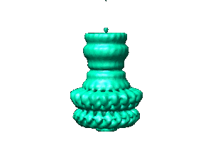 1214.gif 1214.gif | 8.4 KB | ||
| Archive directory |  http://ftp.pdbj.org/pub/emdb/structures/EMD-1214ftp://ftp.pdbj.org/pub/emdb/structures/EMD-1214 http://ftp.pdbj.org/pub/emdb/structures/EMD-1214ftp://ftp.pdbj.org/pub/emdb/structures/EMD-1214 | HTTPS FTP |
-Related structure data


-Links
| EMDB pages | EMDB (EBI/PDBe) / EMDataResource |
|---|
-Map
| File | Download / File: emd_1214.map.gz / Format: CCP4 / Size: 29.8 MB / Type: IMAGE STORED AS FLOATING POINT NUMBER (4 BYTES) | ||||||||||||||||||||||||||||||||||||||||||||||||||||||||||||||||||||
|---|---|---|---|---|---|---|---|---|---|---|---|---|---|---|---|---|---|---|---|---|---|---|---|---|---|---|---|---|---|---|---|---|---|---|---|---|---|---|---|---|---|---|---|---|---|---|---|---|---|---|---|---|---|---|---|---|---|---|---|---|---|---|---|---|---|---|---|---|---|
| Annotation | Type III Secretion Complex from an invJ-mutant strain | ||||||||||||||||||||||||||||||||||||||||||||||||||||||||||||||||||||
| Projections & slices | Image control
Images are generated by Spider. | ||||||||||||||||||||||||||||||||||||||||||||||||||||||||||||||||||||
| Voxel size | X=Y=Z: 2.8 Å | ||||||||||||||||||||||||||||||||||||||||||||||||||||||||||||||||||||
| Density |
| ||||||||||||||||||||||||||||||||||||||||||||||||||||||||||||||||||||
| Symmetry | Space group: 1 | ||||||||||||||||||||||||||||||||||||||||||||||||||||||||||||||||||||
| Details | EMDB XML:
CCP4 map header:
| ||||||||||||||||||||||||||||||||||||||||||||||||||||||||||||||||||||
 Z (Sec.)
 Y (Row.)
 X (Col.)


-Supplemental data
- Sample components
-Entire : Type III Secretion Complex from an invJ-mutant strain of Salmonel...
| Entire | Name: Type III Secretion Complex from an invJ-mutant strain of Salmonella typhimurium |
|---|---|
| Components |
|
-Supramolecule #1000: Type III Secretion Complex from an invJ-mutant strain of Salmonel...
| Supramolecule | Name: Type III Secretion Complex from an invJ-mutant strain of Salmonella typhimurium type: sample / ID: 1000 / Oligomeric state: 20 / Number unique components: 3 |
|---|---|
| Molecular weight | Theoretical: 2.64 MDa |
-Macromolecule #1: PrgH
| Macromolecule | Name: PrgH / type: protein_or_peptide / ID: 1 / Number of copies: 20 / Recombinant expression: Yes |
|---|---|
| Source (natural) | Organism: Salmonella enterica subsp. enterica serovar Typhimurium (bacteria) synonym: Salmonella / Location in cell: Membrane |
| Molecular weight | Experimental: 44 MDa |
| Recombinant expression | Organism: Salmonella enterica subsp. enterica serovar Typhimurium (bacteria) |
-Macromolecule #2: PrgK
| Macromolecule | Name: PrgK / type: protein_or_peptide / ID: 2 / Number of copies: 20 / Recombinant expression: Yes |
|---|---|
| Source (natural) | Organism: Salmonella enterica subsp. enterica serovar Typhimurium (bacteria) synonym: Salmonella / Location in cell: Membrane |
| Molecular weight | Experimental: 28 MDa |
| Recombinant expression | Organism: Salmonella enterica subsp. enterica serovar Typhimurium (bacteria) |
-Macromolecule #3: InvG
| Macromolecule | Name: InvG / type: protein_or_peptide / ID: 3 / Number of copies: 20 / Recombinant expression: Yes |
|---|---|
| Source (natural) | Organism: Salmonella enterica subsp. enterica serovar Typhimurium (bacteria) synonym: Salmonella / Location in cell: Membrane |
| Molecular weight | Experimental: 60 MDa |
| Recombinant expression | Organism: Salmonella enterica subsp. enterica serovar Typhimurium (bacteria) |
-Experimental details
-Structure determination
| Method | cryo EM |
|---|---|
 Processing Processing | single particle reconstruction |
| Aggregation state | particle |
-Sample preparation
| Buffer | pH: 8 / Details: 10mM Tris-HCl 500mM NaCl 0.2% LDAO |
|---|---|
| Grid | Details: 400 mesh Cu/Rh |
| Vitrification | Cryogen name: ETHANE / Method: Blot for 15 seconds before plunging |
- Electron microscopy
| Microscope | FEI TECNAI F20 |
|---|---|
| Alignment procedure | Legacy - Astigmatism: 200 |
| Image recording | Category: FILM / Film or detector model: KODAK SO-163 FILM / Digitization - Scanner: ZEISS SCAI / Digitization - Sampling interval: 7 µm / Number real images: 61 / Average electron dose: 10 e/Å2 / Od range: 0.8 / Bits/pixel: 8 |
| Electron beam | Acceleration voltage: 200 kV / Electron source:  FIELD EMISSION GUN FIELD EMISSION GUN |
| Electron optics | Illumination mode: FLOOD BEAM / Imaging mode: BRIGHT FIELD / Cs: 2.0 mm / Nominal defocus max: 4.2 µm / Nominal defocus min: 1.7 µm / Nominal magnification: 50000 |
| Sample stage | Specimen holder: Side-entry liquid nitrogen-cooled cryo specimen holder Specimen holder model: GATAN LIQUID NITROGEN |
| Experimental equipment |  Model: Tecnai F20 / Image courtesy: FEI Company |
-Image processing
| CTF correction | Details: phase flipping, each particle |
|---|---|
| Final reconstruction | Applied symmetry - Point group: C20 (20 fold cyclic) / Resolution.type: BY AUTHOR / Resolution: 20.0 Å / Resolution method: FSC 0.5 CUT-OFF / Software - Name: SPIDER, IMAGIC-5, MRC / Details: supervised projection matching procedure |
