 Movie
Movie Controller


+ Open data

- Basic information
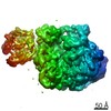
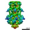
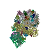
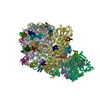
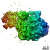
| Entry | Database: PDB / ID: 7a7d | ||||||
|---|---|---|---|---|---|---|---|
| Title | Cadherin fit into cryo-ET map | ||||||
 Components Components |
| ||||||
 Keywords Keywords | CELL ADHESION / Cadherin / Desmosome | ||||||
| Function / homology |  Function and homology information Function and homology informationcardiac muscle cell-cardiac muscle cell adhesion / Purkinje myocyte development / positive regulation of protein localization to cell-cell junction / bundle of His cell-Purkinje myocyte adhesion involved in cell communication / cell adhesive protein binding involved in bundle of His cell-Purkinje myocyte communication / desmosome organization / negative regulation of endothelial cell differentiation / Keratinization / negative regulation of inflammatory response to wounding / desmosome ...cardiac muscle cell-cardiac muscle cell adhesion / Purkinje myocyte development / positive regulation of protein localization to cell-cell junction / bundle of His cell-Purkinje myocyte adhesion involved in cell communication / cell adhesive protein binding involved in bundle of His cell-Purkinje myocyte communication / desmosome organization / negative regulation of endothelial cell differentiation / Keratinization / negative regulation of inflammatory response to wounding / desmosome / mesenchymal to epithelial transition / Formation of the cornified envelope / cornified envelope / Apoptotic cleavage of cell adhesion proteins / regulation of ventricular cardiac muscle cell action potential / positive regulation of p38MAPK cascade / negative regulation of epithelial to mesenchymal transition / positive regulation of sprouting angiogenesis / homophilic cell-cell adhesion / positive regulation of stem cell population maintenance / regulation of heart rate by cardiac conduction / RHOG GTPase cycle / intercalated disc / lateral plasma membrane / RAC3 GTPase cycle / RAC2 GTPase cycle / maternal process involved in female pregnancy / response to progesterone / cell adhesion molecule binding / positive regulation of cell adhesion / stem cell proliferation / cellular response to starvation / adherens junction / cell-cell adhesion / cell-cell junction / cell junction / cytoplasmic vesicle / cell adhesion / apical plasma membrane / calcium ion binding / negative regulation of apoptotic process / cell surface / endoplasmic reticulum / extracellular exosome / plasma membrane / cytoplasm Similarity search - Function | ||||||
| Biological species |  Homo sapiens (human) Homo sapiens (human) | ||||||
| Method | ELECTRON MICROSCOPY / subtomogram averaging / cryo EM / Resolution: 26 Å | ||||||
 Authors Authors | Sikora, M. / Ermel, U.H. / Seybold, A. / Kunz, M. / Calloni, G. / Reitz, J. / Vabulas, R.M. / Hummer, G. / Frangakis, A.S. | ||||||
 Citation Citation | Journal: Proc Natl Acad Sci U S A / Year: 2020 Title: Desmosome architecture derived from molecular dynamics simulations and cryo-electron tomography. Authors: Mateusz Sikora / Utz H Ermel / Anna Seybold / Michael Kunz / Giulia Calloni / Julian Reitz / R Martin Vabulas / Gerhard Hummer / Achilleas S Frangakis /   Abstract: Desmosomes are cell-cell junctions that link tissue cells experiencing intense mechanical stress. Although the structure of the desmosomal cadherins is known, the desmosome architecture-which is ...Desmosomes are cell-cell junctions that link tissue cells experiencing intense mechanical stress. Although the structure of the desmosomal cadherins is known, the desmosome architecture-which is essential for mediating numerous functions-remains elusive. Here, we recorded cryo-electron tomograms (cryo-ET) in which individual cadherins can be discerned; they appear variable in shape, spacing, and tilt with respect to the membrane. The resulting sub-tomogram average reaches a resolution of ∼26 Å, limited by the inherent flexibility of desmosomes. To address this challenge typical of dynamic biological assemblies, we combine sub-tomogram averaging with atomistic molecular dynamics (MD) simulations. We generate models of possible cadherin arrangements and perform an in silico screening according to biophysical and structural properties extracted from MD simulation trajectories. We find a truss-like arrangement of cadherins that resembles the characteristic footprint seen in the electron micrograph. The resulting model of the desmosomal architecture explains their unique biophysical properties and strength. | ||||||
| History |
|
- Structure visualization
| Movie |
Movie viewer |
|---|---|
| Structure viewer | Molecule: MolmilJmol/JSmol |
- Downloads & links
-Download
| PDBx/mmCIF format | 7a7d.cif.gz | 2.2 MB | Display | PDBx/mmCIF format |
|---|---|---|---|---|
| PDB format | pdb7a7d.ent.gz | Display | PDB format | |
| PDBx/mmJSON format | 7a7d.json.gz | Tree view | PDBx/mmJSON format | |
| Others |  Other downloads Other downloads |
-Validation report
| Arichive directory | https://data.pdbj.org/pub/pdb/validation_reports/a7/7a7dftp://data.pdbj.org/pub/pdb/validation_reports/a7/7a7d | HTTPS FTP |
|---|
-Related structure data
| Related structure data |  11678MC M: map data used to model this data C: citing same article ( |
|---|---|
| Similar structure data |

-Links
 PDBj
PDBj


- Assembly
| Deposited unit | 
|
|---|---|
| 1 |
|
-Components
| #1: Protein | Mass: 61893.297 Da / Num. of mol.: 7 / Source method: isolated from a natural source / Source: (natural) Homo sapiens (human) / References: UniProt: Q14126#2: Protein | Mass: 60869.840 Da / Num. of mol.: 7 / Source method: isolated from a natural source / Source: (natural) Homo sapiens (human) / References: UniProt: Q02487Has ligand of interest | N | Has protein modification | Y | |
|---|
-Experimental details
-Experiment
| Experiment | Method: ELECTRON MICROSCOPY |
|---|---|
| EM experiment | Aggregation state: TISSUE / 3D reconstruction method: subtomogram averaging |
- Sample preparation
| Component | Name: Desmosome from mouse liver / Type: ORGANELLE OR CELLULAR COMPONENT / Entity ID: all / Source: NATURAL |
|---|---|
| Source (natural) | Organism: Homo sapiens (human) |
| Buffer solution | pH: 7.5 |
| Specimen | Embedding applied: NO / Shadowing applied: NO / Staining applied: NO / Vitrification applied: YES |
| Vitrification | Cryogen name: ETHANE |
- Electron microscopy imaging
| Experimental equipment |  Model: Titan Krios / Image courtesy: FEI Company |
|---|---|
| Microscopy | Model: FEI TITAN KRIOS |
| Electron gun | Electron source:  FIELD EMISSION GUN / Accelerating voltage: 300 kV / Illumination mode: FLOOD BEAM FIELD EMISSION GUN / Accelerating voltage: 300 kV / Illumination mode: FLOOD BEAM |
| Electron lens | Mode: BRIGHT FIELD / Nominal magnification: 64000 X / Cs: 2.7 mm |
| Specimen holder | Cryogen: NITROGEN / Specimen holder model: FEI TITAN KRIOS AUTOGRID HOLDER |
| Image recording | Average exposure time: 2 sec. / Electron dose: 1.95 e/Å2 / Detector mode: SUPER-RESOLUTION / Film or detector model: GATAN K2 SUMMIT (4k x 4k) |
| EM imaging optics | Energyfilter name: GIF Quantum SE |
| Image scans | Movie frames/image: 4 |
- Processing
| EM software | Name: SerialEM / Category: image acquisition |
|---|---|
| CTF correction | Type: PHASE FLIPPING AND AMPLITUDE CORRECTION |
| Symmetry | Point symmetry: C1 (asymmetric) |
| 3D reconstruction | Resolution: 26 Å / Resolution method: FSC 0.5 CUT-OFF / Num. of particles: 3656 / Symmetry type: POINT |
| EM volume selection | Num. of tomograms: 12 / Num. of volumes extracted: 9000 |
