 Movie
Movie Controller


+ Open data

- Basic information
| Entry | Database: EMDB / ID: EMD-3560 | |||||||||
|---|---|---|---|---|---|---|---|---|---|---|
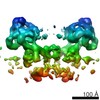
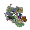
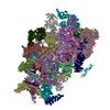
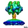
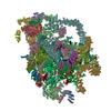
| Title | mitochondrial ATP synthase dimer from Trypanosoma brucei | |||||||||
 Map data Map data | subtomogram average of mitochondrial ATP synthase dimer from Trypanosoma brucei | |||||||||
 Sample Sample |
| |||||||||
| Biological species |  | |||||||||
| Method | subtomogram averaging / cryo EM / Resolution: 32.5 Å | |||||||||
 Authors Authors | Muehleip AW / Kuehlbrandt W / Davies KM | |||||||||
 Citation Citation | Journal: Proc Natl Acad Sci U S A / Year: 2017 Title: In situ structure of trypanosomal ATP synthase dimer reveals a unique arrangement of catalytic subunits. Authors: Alexander W Mühleip / Caroline E Dewar / Achim Schnaufer / Werner Kühlbrandt / Karen M Davies /   Abstract: We used electron cryotomography and subtomogram averaging to determine the in situ structures of mitochondrial ATP synthase dimers from two organisms belonging to the phylum euglenozoa: Trypanosoma ...We used electron cryotomography and subtomogram averaging to determine the in situ structures of mitochondrial ATP synthase dimers from two organisms belonging to the phylum euglenozoa: Trypanosoma brucei, a lethal human parasite, and Euglena gracilis, a photosynthetic protist. At a resolution of 32.5 Å and 27.5 Å, respectively, the two structures clearly exhibit a noncanonical F head, in which the catalytic (αβ) assembly forms a triangular pyramid rather than the pseudo-sixfold ring arrangement typical of all other ATP synthases investigated so far. Fitting of known X-ray structures reveals that this unusual geometry results from a phylum-specific cleavage of the α subunit, in which the C-terminal α fragments are displaced by ∼20 Å and rotated by ∼30° from their expected positions. In this location, the α fragment is unable to form the conserved catalytic interface that was thought to be essential for ATP synthesis, and cannot convert γ-subunit rotation into the conformational changes implicit in rotary catalysis. The new arrangement of catalytic subunits suggests that the mechanism of ATP generation by rotary ATPases is less strictly conserved than has been generally assumed. The ATP synthases of these organisms present a unique model system for discerning the individual contributions of the α and β subunits to the fundamental process of ATP synthesis. | |||||||||
| History |
|
- Structure visualization
| Movie |
 Movie viewer Movie viewer |
|---|---|
| Structure viewer | EM map: SurfViewMolmilJmol/JSmol |
| Supplemental images |

- Downloads & links
-EMDB archive
| Map data | emd_3560.map.gz | 3.7 MB | EMDB map data format | |
|---|---|---|---|---|
| Header (meta data) | emd-3560-v30.xmlemd-3560.xml | 11.5 KB 11.5 KB | Display Display | EMDB header |
| Images |  emd_3560.png emd_3560.png | 80.4 KB | ||
| Archive directory |  http://ftp.pdbj.org/pub/emdb/structures/EMD-3560ftp://ftp.pdbj.org/pub/emdb/structures/EMD-3560 http://ftp.pdbj.org/pub/emdb/structures/EMD-3560ftp://ftp.pdbj.org/pub/emdb/structures/EMD-3560 | HTTPS FTP |
-Related structure data
-Links
| EMDB pages | EMDB (EBI/PDBe) / EMDataResource |
|---|
-Map
| File | Download / File: emd_3560.map.gz / Format: CCP4 / Size: 5.1 MB / Type: IMAGE STORED AS FLOATING POINT NUMBER (4 BYTES) | ||||||||||||||||||||||||||||||||||||||||||||||||||||||||||||
|---|---|---|---|---|---|---|---|---|---|---|---|---|---|---|---|---|---|---|---|---|---|---|---|---|---|---|---|---|---|---|---|---|---|---|---|---|---|---|---|---|---|---|---|---|---|---|---|---|---|---|---|---|---|---|---|---|---|---|---|---|---|
| Annotation | subtomogram average of mitochondrial ATP synthase dimer from Trypanosoma brucei | ||||||||||||||||||||||||||||||||||||||||||||||||||||||||||||
| Projections & slices | Image control
Images are generated by Spider. | ||||||||||||||||||||||||||||||||||||||||||||||||||||||||||||
| Voxel size | X=Y=Z: 6.693 Å | ||||||||||||||||||||||||||||||||||||||||||||||||||||||||||||
| Density |
| ||||||||||||||||||||||||||||||||||||||||||||||||||||||||||||
| Symmetry | Space group: 1 | ||||||||||||||||||||||||||||||||||||||||||||||||||||||||||||
| Details | EMDB XML:
CCP4 map header:
| ||||||||||||||||||||||||||||||||||||||||||||||||||||||||||||
 Z (Sec.)
 Y (Row.)
 X (Col.)


-Supplemental data
- Sample components
-Entire : ATP synthase dimer from Trypanosoma brucei in isolated mitochondr...
| Entire | Name: ATP synthase dimer from Trypanosoma brucei in isolated mitochondrial membranes |
|---|---|
| Components |
|
-Supramolecule #1: ATP synthase dimer from Trypanosoma brucei in isolated mitochondr...
| Supramolecule | Name: ATP synthase dimer from Trypanosoma brucei in isolated mitochondrial membranes type: organelle_or_cellular_component / ID: 1 / Parent: 0 |
|---|---|
| Source (natural) | Organism: |
| Molecular weight | Theoretical: 2 MDa |
-Experimental details
-Structure determination
| Method | cryo EM |
|---|---|
 Processing Processing | subtomogram averaging |
| Aggregation state | 3D array |
-Sample preparation
| Buffer | pH: 7.4 Component:
| |||||||||
|---|---|---|---|---|---|---|---|---|---|---|
| Grid | Model: Quantifoil R2/2 / Material: COPPER / Mesh: 300 | |||||||||
| Vitrification | Cryogen name: ETHANE / Chamber humidity: 100 % / Instrument: HOMEMADE PLUNGER |
- Electron microscopy
| Microscope | FEI TITAN KRIOS |
|---|---|
| Temperature | Min: 70.0 K |
| Specialist optics | Energy filter - Name: GIF Quantum / Energy filter - Lower energy threshold: 0 eV / Energy filter - Upper energy threshold: 20 eV |
| Image recording | Film or detector model: GATAN K2 SUMMIT (4k x 4k) / Detector mode: COUNTING / Digitization - Dimensions - Width: 3710 pixel / Digitization - Dimensions - Height: 3838 pixel / Average electron dose: 1.6 e/Å2 |
| Electron beam | Acceleration voltage: 300 kV / Electron source:  FIELD EMISSION GUN FIELD EMISSION GUN |
| Electron optics | C2 aperture diameter: 70.0 µm / Illumination mode: FLOOD BEAM / Imaging mode: BRIGHT FIELD / Cs: 2.7 mm / Nominal defocus max: 4.0 µm / Nominal defocus min: 3.5 µm / Nominal magnification: 42000 |
| Sample stage | Cooling holder cryogen: NITROGEN |
| Experimental equipment |  Model: Titan Krios / Image courtesy: FEI Company |
-Image processing
| Final reconstruction | Applied symmetry - Point group: C1 (asymmetric) / Resolution.type: BY AUTHOR / Resolution: 32.5 Å / Resolution method: FSC 0.5 CUT-OFF / Software: (Name: IMOD, PEET) / Number subtomograms used: 925 |
|---|---|
| Extraction | Number tomograms: 6 / Number images used: 925 / Reference model: crude average from original data / Method: volumes picked interactively / Software: (Name: 3dmod, IMOD) |
| CTF correction | Software - Name: IMOD / Details: strip-based method |
| Final angle assignment | Type: OTHER / Software: (Name: IMOD, PEET) |
-Atomic model buiding 1
| Refinement | Protocol: RIGID BODY FIT |
|---|
