 ムービー
ムービー コントローラー


+ データを開く

- 基本情報
基本情報
| 登録情報 | データベース: EMDB / ID: EMD-3318 | |||||||||
|---|---|---|---|---|---|---|---|---|---|---|
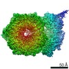
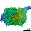
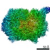
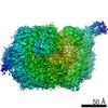
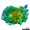
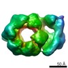
| タイトル | CryoEM structure of the CMG replicative helicase bound to a DNA fork (compact state) | |||||||||
 マップデータ マップデータ | CMG treated with forked DNA substrate in the presence of ATPgS in a compact Mcm5-2 AAA+ configuration | |||||||||
 試料 試料 |
| |||||||||
 キーワード キーワード | Cdc45 / GINS / MCM / CMG / helicase / DNA replication | |||||||||
| 機能・相同性 |  機能・相同性情報 機能・相同性情報Unwinding of DNA / Assembly of the pre-replicative complex / Switching of origins to a post-replicative state / DNA endoreduplication / Activation of ATR in response to replication stress / Activation of the pre-replicative complex / eggshell chorion gene amplification / Orc1 removal from chromatin / DNA amplification / DNA strand elongation involved in mitotic DNA replication ...Unwinding of DNA / Assembly of the pre-replicative complex / Switching of origins to a post-replicative state / DNA endoreduplication / Activation of ATR in response to replication stress / Activation of the pre-replicative complex / eggshell chorion gene amplification / Orc1 removal from chromatin / DNA amplification / DNA strand elongation involved in mitotic DNA replication / GINS complex / mitotic DNA replication preinitiation complex assembly / resolution of meiotic recombination intermediates / premeiotic DNA replication / mitotic DNA replication / CMG complex / DNA replication preinitiation complex / MCM complex / double-strand break repair via break-induced replication / mitotic DNA replication initiation / chromosome condensation / DNA strand elongation involved in DNA replication / DNA replication origin binding / DNA replication initiation / regulation of DNA-templated transcription elongation / mitotic spindle organization / meiotic cell cycle / mitotic cell cycle / single-stranded DNA binding / DNA helicase / forked DNA-dependent helicase activity / single-stranded 3'-5' DNA helicase activity / four-way junction helicase activity / double-stranded DNA helicase activity / DNA replication / cell division / chromatin binding / ATP hydrolysis activity / zinc ion binding / ATP binding / nucleus / cytosol / cytoplasm 類似検索 - 分子機能 | |||||||||
| 生物種 |  | |||||||||
| 手法 | 単粒子再構成法 / クライオ電子顕微鏡法 / 解像度: 7.4 Å | |||||||||
 データ登録者 データ登録者 | Abid Ali F / Renault L / Costa A | |||||||||
 引用 引用 | ジャーナル: Nat Commun / 年: 2016 タイトル: Cryo-EM structures of the eukaryotic replicative helicase bound to a translocation substrate. 著者: Ferdos Abid Ali / Ludovic Renault / Julian Gannon / Hailey L Gahlon / Abhay Kotecha / Jin Chuan Zhou / David Rueda / Alessandro Costa /  要旨: The Cdc45-MCM-GINS (CMG) helicase unwinds DNA during the elongation step of eukaryotic genome duplication and this process depends on the MCM ATPase function. Whether CMG translocation occurs on ...The Cdc45-MCM-GINS (CMG) helicase unwinds DNA during the elongation step of eukaryotic genome duplication and this process depends on the MCM ATPase function. Whether CMG translocation occurs on single- or double-stranded DNA and how ATP hydrolysis drives DNA unwinding remain open questions. Here we use cryo-electron microscopy to describe two subnanometre resolution structures of the CMG helicase trapped on a DNA fork. In the predominant state, the ring-shaped C-terminal ATPase of MCM is compact and contacts single-stranded DNA, via a set of pre-sensor 1 hairpins that spiral around the translocation substrate. In the second state, the ATPase module is relaxed and apparently substrate free, while DNA intimately contacts the downstream amino-terminal tier of the MCM motor ring. These results, supported by single-molecule FRET measurements, lead us to suggest a replication fork unwinding mechanism whereby the N-terminal and AAA+ tiers of the MCM work in concert to translocate on single-stranded DNA. | |||||||||
| 履歴 |
|
- 構造の表示
構造の表示
| ムービー |
ムービービューア |
|---|---|
| 構造ビューア | EMマップ: SurfViewMolmilJmol/JSmol |
| 添付画像 |

- ダウンロードとリンク
-EMDBアーカイブ
| マップデータ | emd_3318.map.gz | 810.4 KB | EMDBマップデータ形式 | |
|---|---|---|---|---|
| ヘッダ (付随情報) | emd-3318-v30.xmlemd-3318.xml | 23.9 KB 23.9 KB | 表示 表示 | EMDBヘッダ |
| FSC (解像度算出) | emd_3318_fsc.xml | 3.6 KB | 表示 | FSCデータファイル |
| 画像 | 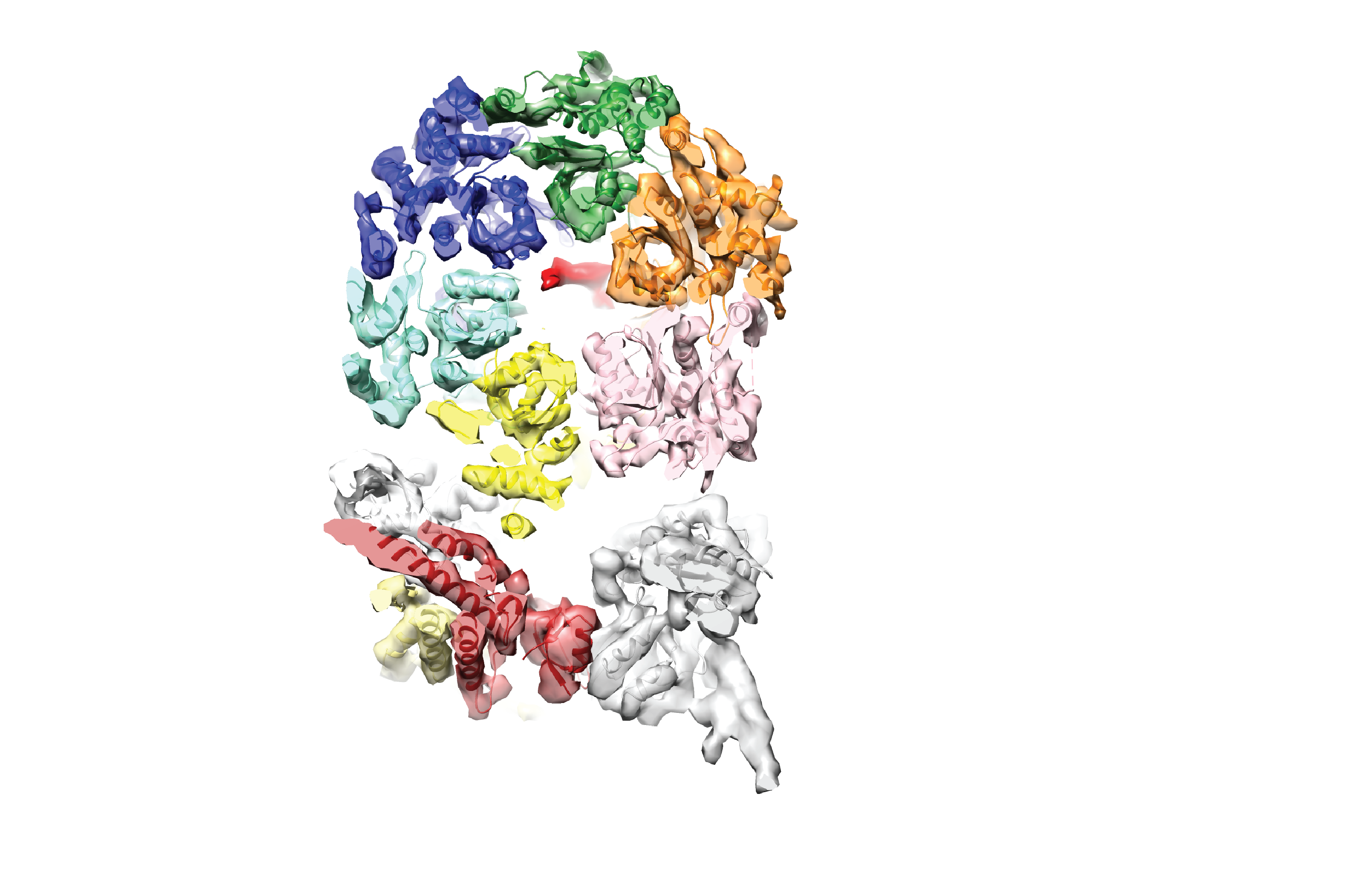 emd_3318.png emd_3318.png | 1.4 MB | ||
| アーカイブディレクトリ |  http://ftp.pdbj.org/pub/emdb/structures/EMD-3318ftp://ftp.pdbj.org/pub/emdb/structures/EMD-3318 http://ftp.pdbj.org/pub/emdb/structures/EMD-3318ftp://ftp.pdbj.org/pub/emdb/structures/EMD-3318 | HTTPS FTP |
-関連構造データ

-リンク
| EMDBのページ | EMDB (EBI/PDBe) / EMDataResource |
|---|---|
| 「今月の分子」の関連する項目 |

-マップ
| ファイル | ダウンロード / ファイル: emd_3318.map.gz / 形式: CCP4 / 大きさ: 5.2 MB / タイプ: IMAGE STORED AS FLOATING POINT NUMBER (4 BYTES) | ||||||||||||||||||||||||||||||||||||||||||||||||||||||||||||
|---|---|---|---|---|---|---|---|---|---|---|---|---|---|---|---|---|---|---|---|---|---|---|---|---|---|---|---|---|---|---|---|---|---|---|---|---|---|---|---|---|---|---|---|---|---|---|---|---|---|---|---|---|---|---|---|---|---|---|---|---|---|
| 注釈 | CMG treated with forked DNA substrate in the presence of ATPgS in a compact Mcm5-2 AAA+ configuration | ||||||||||||||||||||||||||||||||||||||||||||||||||||||||||||
| 投影像・断面図 | 画像のコントロール
画像は Spider により作成 | ||||||||||||||||||||||||||||||||||||||||||||||||||||||||||||
| ボクセルのサイズ | X=Y=Z: 2.7 Å | ||||||||||||||||||||||||||||||||||||||||||||||||||||||||||||
| 密度 |
| ||||||||||||||||||||||||||||||||||||||||||||||||||||||||||||
| 対称性 | 空間群: 1 | ||||||||||||||||||||||||||||||||||||||||||||||||||||||||||||
| 詳細 | EMDB XML:
CCP4マップ ヘッダ情報:
| ||||||||||||||||||||||||||||||||||||||||||||||||||||||||||||
 Z (Sec.)
 Y (Row.)
 X (Col.)


-添付データ
- 試料の構成要素
+全体 : CMG bound to DNA fork and ATPgS treated
+超分子 #1000: CMG bound to DNA fork and ATPgS treated
+分子 #1: Mcm2
+分子 #4: Mcm3
 unidentified baculovirus (ウイルス)
+分子 #5: Mcm4
+分子 #6: Mcm5
+分子 #7: Mcm6
+分子 #8: Mcm7
+分子 #9: Cdc45
+分子 #10: Psf1
+分子 #11: Psf2
+分子 #12: Psf3
+分子 #13: Sld5
+分子 #2: Model replication DNA fork leading strand template
+分子 #3: Model replication DNA fork lagging strand template
-実験情報
-構造解析
| 手法 | クライオ電子顕微鏡法 |
|---|---|
 解析 解析 | 単粒子再構成法 |
| 試料の集合状態 | particle |
-試料調製
| 緩衝液 | pH: 7.6 詳細: 25 mM Hepes, 50 mM sodium acetate, 10 mM magnesium acetate, 1 mM DTT, 0.1mM ATPgammaS |
|---|---|
| グリッド | 詳細: Quantifoil 1.2/1.3 or C-flat 1/1 |
| 凍結 | 凍結剤: ETHANE / チャンバー内湿度: 100 % / 装置: FEI VITROBOT MARK IV / 手法: Blot for 4 seconds before plunging |
- 電子顕微鏡法 #1
| Microscopy ID | 1 |
|---|---|
| 顕微鏡 | FEI POLARA 300 |
| 特殊光学系 | エネルギーフィルター - 名称: GIF エネルギーフィルター - エネルギー下限: 0.0 eV エネルギーフィルター - エネルギー上限: 20.0 eV |
| 日付 | 2015年8月7日 |
| 撮影 | カテゴリ: CCD フィルム・検出器のモデル: GATAN K2 SUMMIT (4k x 4k) デジタル化 - サンプリング間隔: 5 µm / 実像数: 2098 / 平均電子線量: 48 e/Å2 / 詳細: data was recorded as movies of 25 frames / ビット/ピクセル: 32 |
| 電子線 | 加速電圧: 300 kV / 電子線源:  FIELD EMISSION GUN FIELD EMISSION GUN |
| 電子光学系 | 照射モード: FLOOD BEAM / 撮影モード: BRIGHT FIELD / Cs: 2.0 mm / 最大 デフォーカス(公称値): 3.5 µm / 最小 デフォーカス(公称値): 1.7 µm / 倍率(公称値): 37037 |
| 試料ステージ | 試料ホルダー: Multi cartridge holder / 試料ホルダーモデル: OTHER |
| 実験機器 |  モデル: Tecnai Polara / 画像提供: FEI Company |
-電子顕微鏡法 #2
| Microscopy ID | 2 |
|---|---|
| 顕微鏡 | FEI POLARA 300 |
| 特殊光学系 | エネルギーフィルター - 名称: GIF エネルギーフィルター - エネルギー下限: 0.0 eV エネルギーフィルター - エネルギー上限: 20.0 eV |
| 日付 | 2015年8月7日 |
| 撮影 | カテゴリ: CCD フィルム・検出器のモデル: GATAN K2 SUMMIT (4k x 4k) デジタル化 - サンプリング間隔: 5 µm / 実像数: 2098 / 平均電子線量: 48 e/Å2 / 詳細: data was recorded as movies of 25 frames / ビット/ピクセル: 32 |
| 電子線 | 加速電圧: 300 kV / 電子線源: FIELD EMISSION GUN |
| 電子光学系 | 照射モード: FLOOD BEAM / 撮影モード: BRIGHT FIELD / Cs: 2.0 mm / 最大 デフォーカス(公称値): 3.5 µm / 最小 デフォーカス(公称値): 1.7 µm / 倍率(公称値): 37037 |
| 試料ステージ | 試料ホルダー: Multi cartridge holder / 試料ホルダーモデル: OTHER |
| 実験機器 | モデル: Tecnai Polara / 画像提供: FEI Company |
-画像解析
| 詳細 | Particles were picked in EMAN2; Contrast Transfer Function was estimated using CTFFIND4. All further processing was performed within the RELION 1.4 environment. |
|---|---|
| CTF補正 | 詳細: Each particle |
| 最終 再構成 | 想定した対称性 - 点群: C1 (非対称) / 解像度のタイプ: BY AUTHOR / 解像度: 7.4 Å / 解像度の算出法: OTHER / ソフトウェア - 名称: RELION1.4 / 使用した粒子像数: 38792 |
| FSC曲線 (解像度の算出) | 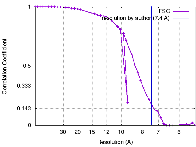 |
