 ムービー
ムービー コントローラー


+ データを開く

- 基本情報
基本情報
| 登録情報 | データベース: EMDB / ID: EMD-2572 | |||||||||
|---|---|---|---|---|---|---|---|---|---|---|
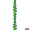
| タイトル | Cryo-EM structure of Plasmodium falciparum actin I | |||||||||
 マップデータ マップデータ | Cryo-EM structure of Plasmodium falciparum actin I | |||||||||
 試料 試料 |
| |||||||||
 キーワード キーワード | Plasmodium falciparum Actin I /  malaria parasite (マラリア原虫) malaria parasite (マラリア原虫) | |||||||||
| 機能・相同性 |  機能・相同性情報 機能・相同性情報Actins signature 1. / Actin, conserved site / Actins signature 2. / Actin/actin-like conserved site / Actins and actin-related proteins signature. / アクチン / Actin family / アクチン / ATPase, nucleotide binding domain類似検索 - ドメイン・相同性 | |||||||||
| 生物種 |  Plasmodium falciparum (マラリア病原虫) Plasmodium falciparum (マラリア病原虫) | |||||||||
| 手法 | 単粒子再構成法 / クライオ電子顕微鏡法 / 解像度: 25.0 Å | |||||||||
 データ登録者 データ登録者 | Vahokoski J / Bhargav SP / Desfosses A / Andreadaki M / Kumpula E / Ignatev A / Martinez SM / Lepper S / Frischknecht F / Siden-Kiamos I ...Vahokoski J / Bhargav SP / Desfosses A / Andreadaki M / Kumpula E / Ignatev A / Martinez SM / Lepper S / Frischknecht F / Siden-Kiamos I / Sachse C / Kursula I | |||||||||
 引用 引用 | ジャーナル: PLoS Pathog / 年: 2014 タイトル: Structural differences explain diverse functions of Plasmodium actins. 著者: Juha Vahokoski / Saligram Prabhakar Bhargav / Ambroise Desfosses / Maria Andreadaki / Esa-Pekka Kumpula / Silvia Muñico Martinez / Alexander Ignatev / Simone Lepper / Friedrich Frischknecht ...著者: Juha Vahokoski / Saligram Prabhakar Bhargav / Ambroise Desfosses / Maria Andreadaki / Esa-Pekka Kumpula / Silvia Muñico Martinez / Alexander Ignatev / Simone Lepper / Friedrich Frischknecht / Inga Sidén-Kiamos / Carsten Sachse / Inari Kursula /    要旨: Actins are highly conserved proteins and key players in central processes in all eukaryotic cells. The two actins of the malaria parasite are among the most divergent eukaryotic actins and also ...Actins are highly conserved proteins and key players in central processes in all eukaryotic cells. The two actins of the malaria parasite are among the most divergent eukaryotic actins and also differ from each other more than isoforms in any other species. Microfilaments have not been directly observed in Plasmodium and are presumed to be short and highly dynamic. We show that actin I cannot complement actin II in male gametogenesis, suggesting critical structural differences. Cryo-EM reveals that Plasmodium actin I has a unique filament structure, whereas actin II filaments resemble canonical F-actin. Both Plasmodium actins hydrolyze ATP more efficiently than α-actin, and unlike any other actin, both parasite actins rapidly form short oligomers induced by ADP. Crystal structures of both isoforms pinpoint several structural changes in the monomers causing the unique polymerization properties. Inserting the canonical D-loop to Plasmodium actin I leads to the formation of long filaments in vitro. In vivo, this chimera restores gametogenesis in parasites lacking actin II, suggesting that stable filaments are required for exflagellation. Together, these data underline the divergence of eukaryotic actins and demonstrate how structural differences in the monomers translate into filaments with different properties, implying that even eukaryotic actins have faced different evolutionary pressures and followed different paths for developing their polymerization properties. | |||||||||
| 履歴 |
|
- 構造の表示
構造の表示
| ムービー |
ムービービューア |
|---|---|
| 構造ビューア | EMマップ: SurfViewMolmilJmol/JSmol |
| 添付画像 |

- ダウンロードとリンク
-EMDBアーカイブ
| マップデータ | emd_2572.map.gz | 632.7 KB | EMDBマップデータ形式 | |
|---|---|---|---|---|
| ヘッダ (付随情報) | emd-2572-v30.xmlemd-2572.xml | 9.9 KB 9.9 KB | 表示 表示 | EMDBヘッダ |
| 画像 | 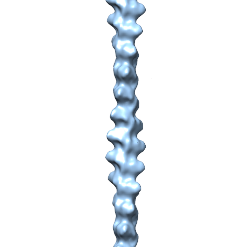 EMD-2572-Actin-I_for-EMDB.png EMD-2572-Actin-I_for-EMDB.png | 51 KB | ||
| アーカイブディレクトリ |  http://ftp.pdbj.org/pub/emdb/structures/EMD-2572ftp://ftp.pdbj.org/pub/emdb/structures/EMD-2572 http://ftp.pdbj.org/pub/emdb/structures/EMD-2572ftp://ftp.pdbj.org/pub/emdb/structures/EMD-2572 | HTTPS FTP |
-関連構造データ

-リンク
| EMDBのページ | EMDB (EBI/PDBe) / EMDataResource |
|---|---|
| 「今月の分子」の関連する項目 |


-マップ
| ファイル | ダウンロード / ファイル: emd_2572.map.gz / 形式: CCP4 / 大きさ: 917 KB / タイプ: IMAGE STORED AS FLOATING POINT NUMBER (4 BYTES) | ||||||||||||||||||||||||||||||||||||||||||||||||||||||||||||||||||||
|---|---|---|---|---|---|---|---|---|---|---|---|---|---|---|---|---|---|---|---|---|---|---|---|---|---|---|---|---|---|---|---|---|---|---|---|---|---|---|---|---|---|---|---|---|---|---|---|---|---|---|---|---|---|---|---|---|---|---|---|---|---|---|---|---|---|---|---|---|---|
| 注釈 | Cryo-EM structure of Plasmodium falciparum actin I | ||||||||||||||||||||||||||||||||||||||||||||||||||||||||||||||||||||
| ボクセルのサイズ | X=Y=Z: 4.412 Å | ||||||||||||||||||||||||||||||||||||||||||||||||||||||||||||||||||||
| 密度 |
| ||||||||||||||||||||||||||||||||||||||||||||||||||||||||||||||||||||
| 対称性 | 空間群: 1 | ||||||||||||||||||||||||||||||||||||||||||||||||||||||||||||||||||||
| 詳細 | EMDB XML:
CCP4マップ ヘッダ情報:
| ||||||||||||||||||||||||||||||||||||||||||||||||||||||||||||||||||||
-添付データ
- 試料の構成要素
-全体 : Plasmodium falciparum Actin I
| 全体 | 名称: Plasmodium falciparum Actin I |
|---|---|
| 要素 |
|
-超分子 #1000: Plasmodium falciparum Actin I
| 超分子 | 名称: Plasmodium falciparum Actin I / タイプ: sample / ID: 1000 / Number unique components: 1 |
|---|
-分子 #1: Plasmodium actin I
| 分子 | 名称: Plasmodium actin I / タイプ: protein_or_peptide / ID: 1 / 組換発現: Yes |
|---|---|
| 由来(天然) | 生物種: Plasmodium falciparum (マラリア病原虫) |
| 組換発現 | 生物種:   Spodoptera frugiperda (ツマジロクサヨトウ) Spodoptera frugiperda (ツマジロクサヨトウ)組換株: Sf21 |
| 配列 | UniProtKB: Actin I |
-実験情報
-構造解析
| 手法 | クライオ電子顕微鏡法 |
|---|---|
 解析 解析 | 単粒子再構成法 |
| 試料の集合状態 | helical array |
-試料調製
| 濃度 | 0.3 mg/mL |
|---|---|
| 緩衝液 | 詳細: 50 mM (pH 8.0) Tris-HCl, 500 mM KCl, 20 mM MgCl2, 50 mM DTT, and 10 mM ATP, JAS 5 uM |
| グリッド | 詳細: glow-discharged holey carbon grids (Quantifoil R 2/2) |
| 凍結 | 凍結剤: ETHANE / チャンバー内湿度: 70 % / チャンバー内温度: 120 K / 装置: LEICA EM GP 手法: Polymerized samples were applied in 3-uL aliquots onto freshly glow-discharged holey carbon grids (Quantifoil R 2/2) at 295 K and 70% humidity and vitrified in liquid ethane using a Leica EM ...手法: Polymerized samples were applied in 3-uL aliquots onto freshly glow-discharged holey carbon grids (Quantifoil R 2/2) at 295 K and 70% humidity and vitrified in liquid ethane using a Leica EM GP vitrification robot. |
- 電子顕微鏡法
| 顕微鏡 | FEI TECNAI F20 |
|---|---|
| 電子線 | 加速電圧: 200 kV / 電子線源: FIELD EMISSION GUN |
| 電子光学系 | 倍率(補正後): 69000 / 照射モード: FLOOD BEAM / 撮影モード: BRIGHT FIELDBright-field microscopy / Cs: 2.0 mm / 最大 デフォーカス(公称値): 7.0 µm / 最小 デフォーカス(公称値): 1.0 µm / 倍率(公称値): 69000 |
| 試料ステージ | 試料ホルダー: 93 K / 試料ホルダーモデル: GATAN LIQUID NITROGEN |
| 日付 | 2013年2月6日 |
| 撮影 | カテゴリ: CCD フィルム・検出器のモデル: GATAN ULTRASCAN 4000 (4k x 4k) 実像数: 75 |
| 実験機器 |  モデル: Tecnai F20 / 画像提供: FEI Company |
-画像解析
| CTF補正 | 詳細: Each particle (convolved) |
|---|---|
| 最終 再構成 | 想定した対称性 - らせんパラメータ - Δz: 27.69 Å 想定した対称性 - らせんパラメータ - ΔΦ: 167.52 ° 想定した対称性 - らせんパラメータ - 軸対称性: C1 (非対称) 解像度のタイプ: BY AUTHOR / 解像度: 25.0 Å / 解像度の算出法: OTHER / ソフトウェア - 名称: SPRING / 使用した粒子像数: 3513 |
| 詳細 | For 3D structure determination, segments were excised using a regular step size of 70 Angstrom, convolved by their respective CTF and further reconstructed as described (Sachse et al. 2007) using the software SPRING (Desfosses et al. 2014) |
