 Movie
Movie Controller


+ Open data

- Basic information
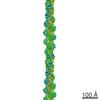
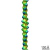
| Entry | Database: PDB / ID: 5mva | |||||||||||||||
|---|---|---|---|---|---|---|---|---|---|---|---|---|---|---|---|---|
| Title | Structure of the thin filament at high calcium concentration | |||||||||||||||
 Components Components | Actin, alpha skeletal muscle | |||||||||||||||
 Keywords Keywords | STRUCTURAL PROTEIN / Thin filament / troponin / actin / tropomyosin | |||||||||||||||
| Function / homology |  Function and homology information Function and homology informationcytoskeletal motor activator activity / myosin heavy chain binding / tropomyosin binding / actin filament bundle / troponin I binding / filamentous actin / mesenchyme migration / skeletal muscle myofibril / actin filament bundle assembly / striated muscle thin filament ...cytoskeletal motor activator activity / myosin heavy chain binding / tropomyosin binding / actin filament bundle / troponin I binding / filamentous actin / mesenchyme migration / skeletal muscle myofibril / actin filament bundle assembly / striated muscle thin filament / skeletal muscle thin filament assembly / actin monomer binding / skeletal muscle fiber development / stress fiber / titin binding / actin filament polymerization / filopodium / actin filament / Hydrolases; Acting on acid anhydrides; Acting on acid anhydrides to facilitate cellular and subcellular movement / calcium-dependent protein binding / lamellipodium / cell body / protein domain specific binding / hydrolase activity / calcium ion binding / positive regulation of gene expression / magnesium ion binding / ATP binding / identical protein binding / cytoplasm Similarity search - Function | |||||||||||||||
| Biological species |  | |||||||||||||||
| Method | ELECTRON MICROSCOPY / single particle reconstruction / negative staining / Resolution: 27.7 Å | |||||||||||||||
 Authors Authors | Paul, D.M. / Squire, J.M. / Morris, E.P. | |||||||||||||||
| Funding support |  United Kingdom, 1items United Kingdom, 1items
| |||||||||||||||
 Citation Citation | Journal: J Struct Biol / Year: 2017 Title: Relaxed and active thin filament structures; a new structural basis for the regulatory mechanism. Authors: Danielle M Paul / John M Squire / Edward P Morris / Abstract: The structures of muscle thin filaments reconstituted using skeletal actin and cardiac troponin and tropomyosin have been determined with and without bound Ca using electron microscopy and reference- ...The structures of muscle thin filaments reconstituted using skeletal actin and cardiac troponin and tropomyosin have been determined with and without bound Ca using electron microscopy and reference-free single particle analysis. The resulting density maps have been fitted with atomic models of actin, tropomyosin and troponin showing that: (i) the polarity of the troponin complex is consistent with our 2009 findings, with large shape changes in troponin between the two states; (ii) without Ca the tropomyosin pseudo-repeats all lie at almost equivalent positions in the 'blocked' position on actin (over subdomains 1 and 2); (iii) in the active state the tropomyosin pseudo-repeats are all displaced towards subdomains 3 and 4 of actin, but the extent of displacement varies within the regulatory unit depending upon the axial location of the pseudo-repeats with respect to troponin. Individual pseudo-repeats with Ca bound to troponin can be assigned either to the 'closed' state, a partly activated conformation, or the 'M-state', a fully activated conformation which has previously been thought to occur only when myosin heads bind. These results lead to a modified view of the steric blocking model of thin filament regulation in which cooperative activation is governed by troponin-mediated local interactions of the pseudo-repeats of tropomyosin with actin. | |||||||||||||||
| History |
|
- Structure visualization
| Movie |
Movie viewer |
|---|---|
| Structure viewer | Molecule: MolmilJmol/JSmol |
- Downloads & links
-Download
| PDBx/mmCIF format | 5mva.cif.gz | 1.6 MB | Display | PDBx/mmCIF format |
|---|---|---|---|---|
| PDB format | pdb5mva.ent.gz | 1.3 MB | Display | PDB format |
| PDBx/mmJSON format | 5mva.json.gz | Tree view | PDBx/mmJSON format | |
| Others |  Other downloads Other downloads |
-Validation report
| Arichive directory | https://data.pdbj.org/pub/pdb/validation_reports/mv/5mvaftp://data.pdbj.org/pub/pdb/validation_reports/mv/5mva | HTTPS FTP |
|---|
-Related structure data
| Related structure data | 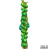 3576MC 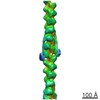 3578C  5mvyC M: map data used to model this data C: citing same article ( |
|---|---|
| Similar structure data |
-Links
 PDBj
PDBj


- Assembly
| Deposited unit | 
|
|---|---|
| 1 |
|
-Components
| #1: Protein | Mass: 41875.633 Da / Num. of mol.: 23 / Source method: isolated from a natural source / Source: (natural) #2: Chemical | ChemComp-ADP /   Mass: 427.201 Da / Num. of mol.: 23 / Source method: isolated from a natural source / Formula: C10H15N5O10P2 / Comment: ADP, energy-carrying molecule*YM Mass: 427.201 Da / Num. of mol.: 23 / Source method: isolated from a natural source / Formula: C10H15N5O10P2 / Comment: ADP, energy-carrying molecule*YMHas protein modification | Y | |
|---|
-Experimental details
-Experiment
| Experiment | Method: ELECTRON MICROSCOPY |
|---|---|
| EM experiment | Aggregation state: FILAMENT / 3D reconstruction method: single particle reconstruction |
- Sample preparation
| Component | Name: Thin filament at high calcium concentration / Type: COMPLEX / Entity ID: #1 / Source: NATURAL |
|---|---|
| Source (natural) | Organism: |
| Buffer solution | pH: 7 |
| Specimen | Embedding applied: NO / Shadowing applied: NO / Staining applied: YES / Vitrification applied: NO |
| EM staining | Type: NEGATIVE / Material: Uranyl Acetate |
- Electron microscopy imaging
| Microscopy | Model: FEI/PHILIPS CM12 |
|---|---|
| Electron gun | Electron source: LAB6 / Accelerating voltage: 120 kV / Illumination mode: FLOOD BEAM |
| Electron lens | Mode: BRIGHT FIELD |
| Image recording | Electron dose: 12 e/Å2 / Film or detector model: AGFA SCIENTA FILM |
| Image scans | Scanner model: ZEISS SCAI |
- Processing
| CTF correction | Type: NONE |
|---|---|
| 3D reconstruction | Resolution: 27.7 Å / Resolution method: FSC 0.5 CUT-OFF / Num. of particles: 1680 / Symmetry type: POINT |
