- EMDB-20184: RF1 accommodated 70S complex at 60 ms -
+
データを開く
IDまたはキーワード:
読み込み中...
-
基本情報
登録情報
データベース: EMDB / ID: EMD-20184
タイトル
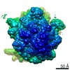
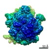
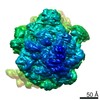
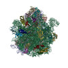
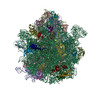
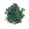
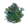
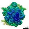
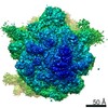
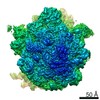
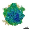
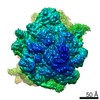
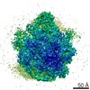
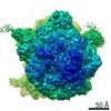
RF1 accommodated 70S complex at 60 ms
マップデータ
RF1o1
試料
複合体: Release complex 70S ribosomes
RNA: x 4種
タンパク質・ペプチド: x 50種
RNA: x 1種
タンパク質・ペプチド: x 1種
リガンド: x 2種
機能・相同性
機能・相同性情報
translation release factor activity, codon specific / ribosomal large subunit assembly / small ribosomal subunit rRNA binding / cytosolic small ribosomal subunit / large ribosomal subunit / cytoplasmic translation / small ribosomal subunit / 5S rRNA binding / cytosolic large ribosomal subunit / transferase activity ...translation release factor activity, codon specific / ribosomal large subunit assembly / small ribosomal subunit rRNA binding / cytosolic small ribosomal subunit / large ribosomal subunit / cytoplasmic translation / small ribosomal subunit / 5S rRNA binding / cytosolic large ribosomal subunit / transferase activity / tRNA binding / rRNA binding / リボソーム / structural constituent of ribosome / ribonucleoprotein complex / 翻訳 (生物学) / mRNA binding / metal ion binding / 細胞質基質 / 細胞質 類似検索 - 分子機能
Peptide chain release factor 1 / Peptide chain release factor / PCRF domain / PCRF / Peptide chain release factor class I superfamily / Prokaryotic-type class I peptide chain release factors signature. / Peptide chain release factor class I / RF-1 domain / Ribosomal protein S21, conserved site / Ribosomal protein S21 signature. ...Peptide chain release factor 1 / Peptide chain release factor / PCRF domain / PCRF / Peptide chain release factor class I superfamily / Prokaryotic-type class I peptide chain release factors signature. / Peptide chain release factor class I / RF-1 domain / Ribosomal protein S21, conserved site / Ribosomal protein S21 signature. / Ribosomal protein L25, short-form / Ribosomal protein S14, bacterial/plastid / Ribosomal protein L31 type A / Ribosomal protein S21 superfamily / Ribosomal protein S21 / Ribosomal protein S16, conserved site / Ribosomal protein S16 signature. / Ribosomal protein L31 signature. / Ribosomal protein S21 / Ribosomal protein L31 / Ribosomal protein L31 superfamily / Ribosomal protein L31 / Ribosomal protein L21, conserved site / Ribosomal protein L21 signature. / Ribosomal protein L16 signature 1. / : / Ribosomal protein L6, conserved site / Ribosomal protein L6 signature 1. / Ribosomal protein L16, conserved site / Ribosomal protein L16 signature 2. / Ribosomal protein L17 signature. / Ribosomal protein L9 signature. / Ribosomal protein L9, bacteria/chloroplast / Ribosomal protein L9, C-terminal / Ribosomal protein L9, C-terminal domain / Ribosomal protein L9, C-terminal domain superfamily / Ribosomal L25p family / Ribosomal protein L25 / Ribosomal protein L28/L24 superfamily / Ribosomal protein L36 signature. / Ribosomal protein L25/Gln-tRNA synthetase, N-terminal / Ribosomal protein L25/Gln-tRNA synthetase, anti-codon-binding domain superfamily / Ribosomal protein L9, N-terminal domain superfamily / Ribosomal protein L9 / Ribosomal protein L9, N-terminal / Ribosomal protein L9, N-terminal domain / Ribosomal protein L28 / Ribosomal protein L35, conserved site / Ribosomal protein L35 signature. / Ribosomal protein L33, conserved site / Ribosomal protein L33 signature. / Ribosomal protein L35, non-mitochondrial / Ribosomal protein L5, bacterial-type / Ribosomal protein L6, bacterial-type / Ribosomal protein L18, bacterial-type / Ribosomal protein L19, conserved site / Ribosomal protein L19 signature. / Ribosomal protein L36 / Ribosomal protein L36 superfamily / Ribosomal protein L36 / Ribosomal protein L9/RNase H1, N-terminal / Ribosomal protein L20 signature. / Ribosomal protein S3, bacterial-type / Ribosomal protein S6, conserved site / Ribosomal protein S6 signature. / Ribosomal protein L27, conserved site / Ribosomal protein L27 signature. / Ribosomal protein S7, bacterial/organellar-type / Ribosomal protein S11, bacterial-type / Ribosomal protein S13, bacterial-type / Ribosomal protein S20 / Ribosomal protein S20 superfamily / Ribosomal protein S20 / Ribosomal protein S9, bacterial/plastid / Ribosomal protein S4, bacterial-type / Ribosomal protein L14P, bacterial-type / Ribosomal protein L34, conserved site / Ribosomal protein L34 signature. / 30S ribosomal protein S17 / Ribosomal protein S5, bacterial-type / Ribosomal protein L22, bacterial/chloroplast-type / Ribosomal protein S6, plastid/chloroplast / Ribosomal protein L35 / Ribosomal protein L35 superfamily / Ribosomal protein L2, bacterial/organellar-type / Ribosomal protein L35 / Ribosomal protein S2, bacteria/mitochondria/plastid / Ribosomal L28 family / Ribosomal protein L33 / Ribosomal protein L33 / Ribosomal protein L28/L24 / Ribosomal protein L33 superfamily / Ribosomal protein L30, bacterial-type / Ribosomal protein L16 / Ribosomal protein L18 / Ribosomal L18 of archaea, bacteria, mitoch. and chloroplast / Ribosomal protein S18, conserved site / Ribosomal protein S18 signature. / L28p-like / Ribosomal protein L20 類似検索 - ドメイン・相同性
50S ribosomal protein L2 / 50S ribosomal protein L23 / Large ribosomal subunit protein uL15 / 30S ribosomal protein S5 / 50S ribosomal protein L31 / Small ribosomal subunit protein uS17 / : / Large ribosomal subunit protein bL36 / 50S ribosomal protein L18 / 50S ribosomal protein L32 ...50S ribosomal protein L2 / 50S ribosomal protein L23 / Large ribosomal subunit protein uL15 / 30S ribosomal protein S5 / 50S ribosomal protein L31 / Small ribosomal subunit protein uS17 / : / Large ribosomal subunit protein bL36 / 50S ribosomal protein L18 / 50S ribosomal protein L32 / 50S ribosomal protein L24 / Small ribosomal subunit protein uS13 / Large ribosomal subunit protein uL6 / Large ribosomal subunit protein uL29 / Large ribosomal subunit protein bL17 / 30S ribosomal protein S3 / Small ribosomal subunit protein bS6 / Peptide chain release factor 1 / Large ribosomal subunit protein bL34 / Small ribosomal subunit protein bS16 / Small ribosomal subunit protein uS2 / Large ribosomal subunit protein bL19 / Large ribosomal subunit protein bL25 / Small ribosomal subunit protein uS15 / 50S ribosomal protein L4 / 50S ribosomal protein L22 / 50S ribosomal protein L14 / 50S ribosomal protein L21 / 50S ribosomal protein L13 / 50S ribosomal protein L9 / 30S ribosomal protein S18 / Small ribosomal subunit protein uS8 / 50S ribosomal protein L33 / 50S ribosomal protein L35 / 30S ribosomal protein S20 / 50S ribosomal protein L30 / 30S ribosomal protein S14 / 30S ribosomal protein S10 / 30S ribosomal protein S21 / 30S ribosomal protein S4 / 30S ribosomal protein S12 / Small ribosomal subunit protein uS11 / Large ribosomal subunit protein uL16 / Large ribosomal subunit protein uL3 / 30S ribosomal protein S7 / Large ribosomal subunit protein bL20 / 30S ribosomal protein S9 / Large ribosomal subunit protein bL27 / Large ribosomal subunit protein uL5 / 50S ribosomal protein L28 類似検索 - 構成要素
National Institutes of Health/National Institute of General Medical Sciences (NIH/NIGMS)
GM55440
米国
Swedish Research Council
スウェーデン
National Institutes of Health/National Institute of General Medical Sciences (NIH/NIGMS)
GM29169
米国
引用
ジャーナル: Nat Commun / 年: 2019 タイトル: The structural basis for release-factor activation during translation termination revealed by time-resolved cryogenic electron microscopy. 著者: Ziao Fu / Gabriele Indrisiunaite / Sandip Kaledhonkar / Binita Shah / Ming Sun / Bo Chen / Robert A Grassucci / Måns Ehrenberg / Joachim Frank / 要旨: When the ribosome encounters a stop codon, it recruits a release factor (RF) to hydrolyze the ester bond between the peptide chain and tRNA. RFs have structural motifs that recognize stop codons in ...When the ribosome encounters a stop codon, it recruits a release factor (RF) to hydrolyze the ester bond between the peptide chain and tRNA. RFs have structural motifs that recognize stop codons in the decoding center and a GGQ motif for induction of hydrolysis in the peptidyl transfer center 70 Å away. Surprisingly, free RF2 is compact, with only 20 Å between its codon-reading and GGQ motifs. Cryo-EM showed that ribosome-bound RFs have extended structures, suggesting that RFs are compact when entering the ribosome and then extend their structures upon stop codon recognition. Here we use time-resolved cryo-EM to visualize transient compact forms of RF1 and RF2 at 3.5 and 4 Å resolution, respectively, in the codon-recognizing ribosome complex on the native pathway. About 25% of complexes have RFs in the compact state at 24 ms reaction time, and within 60 ms virtually all ribosome-bound RFs are transformed to their extended forms.
 ムービー
ムービー コントローラー


 データを開く
 基本情報
基本情報 マップデータ
 試料
試料 機能・相同性情報
 ribosomal large subunit assembly / small ribosomal subunit rRNA binding / cytosolic small ribosomal subunit / large ribosomal subunit / cytoplasmic translation / small ribosomal subunit /
 データ登録者
 米国,
米国,  スウェーデン, 3件
  引用
引用 構造の表示
構造の表示
 ダウンロードとリンク
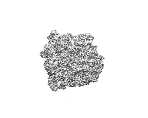 emd_20184.png
 http://ftp.pdbj.org/pub/emdb/structures/EMD-20184


 試料の構成要素
 解析
解析 電子顕微鏡法
