+ Open data
- Basic information
| Entry | Database: PDB / ID: 6osk | |||||||||||||||||||||||||||
|---|---|---|---|---|---|---|---|---|---|---|---|---|---|---|---|---|---|---|---|---|---|---|---|---|---|---|---|---|
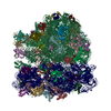
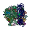
| Title | RF1 accommodated 70S complex at 60 ms | |||||||||||||||||||||||||||
 Components Components |
| |||||||||||||||||||||||||||
 Keywords Keywords | RIBOSOME / Time-resolved cryo-EM / Termination / short-lived / millisecond | |||||||||||||||||||||||||||
| Function / homology |  Function and homology information Function and homology informationtranslation release factor activity, codon specific / large ribosomal subunit / transferase activity / ribosomal large subunit assembly / 5S rRNA binding / small ribosomal subunit / small ribosomal subunit rRNA binding / cytosolic small ribosomal subunit / cytosolic large ribosomal subunit / cytoplasmic translation ...translation release factor activity, codon specific / large ribosomal subunit / transferase activity / ribosomal large subunit assembly / 5S rRNA binding / small ribosomal subunit / small ribosomal subunit rRNA binding / cytosolic small ribosomal subunit / cytosolic large ribosomal subunit / cytoplasmic translation / tRNA binding / rRNA binding / structural constituent of ribosome / ribosome / translation / ribonucleoprotein complex / mRNA binding / metal ion binding / cytoplasm / cytosol Similarity search - Function | |||||||||||||||||||||||||||
| Biological species |  | |||||||||||||||||||||||||||
| Method | ELECTRON MICROSCOPY / single particle reconstruction / cryo EM / Resolution: 3.6 Å | |||||||||||||||||||||||||||
 Authors Authors | Fu, Z. / Indrisiunaite, G. / Kaledhonkar, S. / Shah, B. / Sun, M. / Chen, B. / Grassucci, R.A. / Ehrenberg, M. / Frank, J. | |||||||||||||||||||||||||||
| Funding support |  United States, United States,  Sweden, 3items Sweden, 3items
| |||||||||||||||||||||||||||
 Citation Citation |  Journal: Nat Commun / Year: 2019 Journal: Nat Commun / Year: 2019Title: The structural basis for release-factor activation during translation termination revealed by time-resolved cryogenic electron microscopy. Authors: Ziao Fu / Gabriele Indrisiunaite / Sandip Kaledhonkar / Binita Shah / Ming Sun / Bo Chen / Robert A Grassucci / Måns Ehrenberg / Joachim Frank /   Abstract: When the ribosome encounters a stop codon, it recruits a release factor (RF) to hydrolyze the ester bond between the peptide chain and tRNA. RFs have structural motifs that recognize stop codons in ...When the ribosome encounters a stop codon, it recruits a release factor (RF) to hydrolyze the ester bond between the peptide chain and tRNA. RFs have structural motifs that recognize stop codons in the decoding center and a GGQ motif for induction of hydrolysis in the peptidyl transfer center 70 Å away. Surprisingly, free RF2 is compact, with only 20 Å between its codon-reading and GGQ motifs. Cryo-EM showed that ribosome-bound RFs have extended structures, suggesting that RFs are compact when entering the ribosome and then extend their structures upon stop codon recognition. Here we use time-resolved cryo-EM to visualize transient compact forms of RF1 and RF2 at 3.5 and 4 Å resolution, respectively, in the codon-recognizing ribosome complex on the native pathway. About 25% of complexes have RFs in the compact state at 24 ms reaction time, and within 60 ms virtually all ribosome-bound RFs are transformed to their extended forms. | |||||||||||||||||||||||||||
| History |
|
- Structure visualization
| Movie |
 Movie viewer Movie viewer |
|---|---|
| Structure viewer | Molecule:  Molmil Molmil Jmol/JSmol Jmol/JSmol |
- Downloads & links
- Download
| PDBx/mmCIF format |  6osk.cif.gz 6osk.cif.gz | 3.2 MB | Display |  PDBx/mmCIF format PDBx/mmCIF format |
|---|---|---|---|---|
| PDB format |  pdb6osk.ent.gz pdb6osk.ent.gz | Display |  PDB format PDB format | |
| PDBx/mmJSON format |  6osk.json.gz 6osk.json.gz | Tree view |  PDBx/mmJSON format PDBx/mmJSON format | |
| Others |  Other downloads Other downloads |
-Validation report
| Arichive directory |  https://data.pdbj.org/pub/pdb/validation_reports/os/6osk https://data.pdbj.org/pub/pdb/validation_reports/os/6osk ftp://data.pdbj.org/pub/pdb/validation_reports/os/6osk ftp://data.pdbj.org/pub/pdb/validation_reports/os/6osk | HTTPS FTP |
|---|
-Related structure data
| Related structure data |  20184MC  6oreC  6orlC  6osqC  6ostC  6ot3C  6ouoC M: map data used to model this data C: citing same article ( |
|---|---|
| Similar structure data |
- Links
Links
- Assembly
| Deposited unit | 
|
|---|---|
| 1 |
|
- Components
-RNA chain , 5 types, 5 molecules 12345
| #1: RNA chain | Mass: 941526.438 Da / Num. of mol.: 1 / Source method: isolated from a natural source / Source: (natural)  |
|---|---|
| #2: RNA chain | Mass: 497404.969 Da / Num. of mol.: 1 / Source method: isolated from a natural source / Source: (natural)  |
| #3: RNA chain | Mass: 38790.090 Da / Num. of mol.: 1 / Source method: isolated from a natural source / Source: (natural)  |
| #4: RNA chain | Mass: 2754.647 Da / Num. of mol.: 1 / Source method: isolated from a natural source / Source: (natural)  |
| #55: RNA chain | Mass: 24565.785 Da / Num. of mol.: 1 / Source method: isolated from a natural source / Source: (natural)  |
+50S ribosomal protein ... , 29 types, 29 molecules BCDEFGJKLMNOPQRSTUVWXYZabcdef
-30S ribosomal protein ... , 20 types, 20 molecules ghijklmnopqrstuvwxyz
| #34: Protein | Mass: 25072.867 Da / Num. of mol.: 1 / Source method: isolated from a natural source / Source: (natural)  |
|---|---|
| #35: Protein | Mass: 23248.994 Da / Num. of mol.: 1 / Source method: isolated from a natural source / Source: (natural)  |
| #36: Protein | Mass: 23383.002 Da / Num. of mol.: 1 / Source method: isolated from a natural source / Source: (natural)  |
| #37: Protein | Mass: 16475.037 Da / Num. of mol.: 1 / Source method: isolated from a natural source / Source: (natural) Escherichia coli / References: UniProt: A0A073FR78 |
| #38: Protein | Mass: 12125.993 Da / Num. of mol.: 1 / Source method: isolated from a natural source / Source: (natural)  |
| #39: Protein | Mass: 16861.523 Da / Num. of mol.: 1 / Source method: isolated from a natural source / Source: (natural)  |
| #40: Protein | Mass: 14015.361 Da / Num. of mol.: 1 / Source method: isolated from a natural source / Source: (natural)  |
| #41: Protein | Mass: 14554.882 Da / Num. of mol.: 1 / Source method: isolated from a natural source / Source: (natural)  |
| #42: Protein | Mass: 11254.041 Da / Num. of mol.: 1 / Source method: isolated from a natural source / Source: (natural) Escherichia coli / References: UniProt: J7RLQ6 |
| #43: Protein | Mass: 12487.200 Da / Num. of mol.: 1 / Source method: isolated from a natural source / Source: (natural)  |
| #44: Protein | Mass: 13683.053 Da / Num. of mol.: 1 / Source method: isolated from a natural source / Source: (natural) Escherichia coli / References: UniProt: L4V1L2 |
| #45: Protein | Mass: 12868.091 Da / Num. of mol.: 1 / Source method: isolated from a natural source / Source: (natural) Escherichia coli / References: UniProt: A0A1X3JB38 |
| #46: Protein | Mass: 11475.364 Da / Num. of mol.: 1 / Source method: isolated from a natural source / Source: (natural) Escherichia coli / References: UniProt: J7R6H7 |
| #47: Protein | Mass: 10159.621 Da / Num. of mol.: 1 / Source method: isolated from a natural source / Source: (natural)  |
| #48: Protein | Mass: 9207.572 Da / Num. of mol.: 1 / Source method: isolated from a natural source / Source: (natural)  |
| #49: Protein | Mass: 9263.946 Da / Num. of mol.: 1 / Source method: isolated from a natural source / Source: (natural)  |
| #50: Protein | Mass: 7734.896 Da / Num. of mol.: 1 / Source method: isolated from a natural source / Source: (natural) Escherichia coli / References: UniProt: D7ZI16 |
| #51: Protein | Mass: 9421.018 Da / Num. of mol.: 1 / Source method: isolated from a natural source / Source: (natural) Escherichia coli / References: UniProt: A0A0A8UF41 |
| #52: Protein | Mass: 9577.268 Da / Num. of mol.: 1 / Source method: isolated from a natural source / Source: (natural) Escherichia coli / References: UniProt: I4T5W9 |
| #53: Protein | Mass: 8392.844 Da / Num. of mol.: 1 / Source method: isolated from a natural source / Source: (natural) Escherichia coli / References: UniProt: L3C5D9 |
-Protein / Protein/peptide , 2 types, 2 molecules A6
| #54: Protein | Mass: 28727.230 Da / Num. of mol.: 1 / Source method: isolated from a natural source / Source: (natural)  |
|---|---|
| #56: Protein/peptide | Mass: 471.569 Da / Num. of mol.: 1 / Source method: isolated from a natural source / Source: (natural)  |
-Non-polymers , 2 types, 309 molecules 


| #57: Chemical | ChemComp-MG / #58: Chemical | |
|---|
-Details
| Has protein modification | Y |
|---|
-Experimental details
-Experiment
| Experiment | Method: ELECTRON MICROSCOPY |
|---|---|
| EM experiment | Aggregation state: PARTICLE / 3D reconstruction method: single particle reconstruction |
- Sample preparation
| Component | Name: Release complex 70S ribosomes / Type: RIBOSOME / Entity ID: #1-#54 / Source: NATURAL |
|---|---|
| Molecular weight | Units: MEGADALTONS / Experimental value: NO |
| Source (natural) | Organism:  |
| Buffer solution | pH: 7.4 |
| Specimen | Embedding applied: NO / Shadowing applied: NO / Staining applied: NO / Vitrification applied: YES |
| Specimen support | Details: unspecified |
| Vitrification | Cryogen name: ETHANE-PROPANE |
- Electron microscopy imaging
| Experimental equipment |  Model: Tecnai Polara / Image courtesy: FEI Company |
|---|---|
| Microscopy | Model: FEI POLARA 300 |
| Electron gun | Electron source:  FIELD EMISSION GUN / Accelerating voltage: 300 kV / Illumination mode: FLOOD BEAM FIELD EMISSION GUN / Accelerating voltage: 300 kV / Illumination mode: FLOOD BEAM |
| Electron lens | Mode: BRIGHT FIELD |
| Image recording | Electron dose: 41.6 e/Å2 / Film or detector model: GATAN K2 SUMMIT (4k x 4k) |
- Processing
| CTF correction | Type: PHASE FLIPPING AND AMPLITUDE CORRECTION |
|---|---|
| 3D reconstruction | Resolution: 3.6 Å / Resolution method: FSC 0.143 CUT-OFF / Num. of particles: 202103 / Symmetry type: POINT |
 Movie
Movie Controller





 PDBj
PDBj


