 ムービー
ムービー コントローラー


+ データを開く

- 基本情報
基本情報
| 登録情報 | データベース: EMDB / ID: EMD-1787 | |||||||||
|---|---|---|---|---|---|---|---|---|---|---|
| タイトル | Asymmetric reconstruction of doublecortin-stabilised microtubules decorated with kinesin motor domain | |||||||||
 マップデータ マップデータ | This is a C1 reconstruction of doublecortin-stabilised microtubule decorated with kinesin motor domain | |||||||||
 試料 試料 |
| |||||||||
 キーワード キーワード | Tubulin / MAP / microtubule / stabilisation / doublecortin / kinesin | |||||||||
| 生物種 |   Homo sapiens (ヒト) / Homo sapiens (ヒト) / | |||||||||
| 手法 | 単粒子再構成法 / クライオ電子顕微鏡法 / ネガティブ染色法 / 解像度: 13.5 Å | |||||||||
 データ登録者 データ登録者 | Fourniol FJ / Sindelar CV / Amigues B / Clare D / Thomas G / Perderiset M / Francis F / Houdusse A / Moores CA | |||||||||
 引用 引用 | ジャーナル: J Cell Biol / 年: 2010 タイトル: Template-free 13-protofilament microtubule-MAP assembly visualized at 8 A resolution. 著者: Franck J Fourniol / Charles V Sindelar / Béatrice Amigues / Daniel K Clare / Geraint Thomas / Mylène Perderiset / Fiona Francis / Anne Houdusse / Carolyn A Moores /  要旨: Microtubule-associated proteins (MAPs) are essential for regulating and organizing cellular microtubules (MTs). However, our mechanistic understanding of MAP function is limited by a lack of detailed ...Microtubule-associated proteins (MAPs) are essential for regulating and organizing cellular microtubules (MTs). However, our mechanistic understanding of MAP function is limited by a lack of detailed structural information. Using cryo-electron microscopy and single particle algorithms, we solved the 8 Å structure of doublecortin (DCX)-stabilized MTs. Because of DCX's unusual ability to specifically nucleate and stabilize 13-protofilament MTs, our reconstruction provides unprecedented insight into the structure of MTs with an in vivo architecture, and in the absence of a stabilizing drug. DCX specifically recognizes the corner of four tubulin dimers, a binding mode ideally suited to stabilizing both lateral and longitudinal lattice contacts. A striking consequence of this is that DCX does not bind the MT seam. DCX binding on the MT surface indirectly stabilizes conserved tubulin-tubulin lateral contacts in the MT lumen, operating independently of the nucleotide bound to tubulin. DCX's exquisite binding selectivity uncovers important insights into regulation of cellular MTs. | |||||||||
| 履歴 |
|
- 構造の表示
構造の表示
| ムービー |
 ムービービューア ムービービューア |
|---|---|
| 構造ビューア | EMマップ: SurfViewMolmilJmol/JSmol |
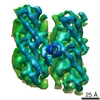
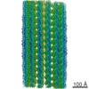
| 添付画像 |

- ダウンロードとリンク
-EMDBアーカイブ
| マップデータ | emd_1787.map.gz | 28 MB | EMDBマップデータ形式 | |
|---|---|---|---|---|
| ヘッダ (付随情報) | emd-1787-v30.xmlemd-1787.xml | 11.6 KB 11.6 KB | 表示 表示 | EMDBヘッダ |
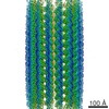
| 画像 | 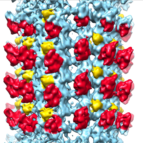 1787_image.png 1787_image.png | 429.2 KB | ||
| アーカイブディレクトリ |  http://ftp.pdbj.org/pub/emdb/structures/EMD-1787ftp://ftp.pdbj.org/pub/emdb/structures/EMD-1787 http://ftp.pdbj.org/pub/emdb/structures/EMD-1787ftp://ftp.pdbj.org/pub/emdb/structures/EMD-1787 | HTTPS FTP |
-検証レポート
| 文書・要旨 | emd_1787_validation.pdf.gz | 263.9 KB | 表示 | EMDB検証レポート |
|---|---|---|---|---|
| 文書・詳細版 | emd_1787_full_validation.pdf.gz | 263 KB | 表示 | |
| XML形式データ | emd_1787_validation.xml.gz | 6.3 KB | 表示 | |
| アーカイブディレクトリ | https://ftp.pdbj.org/pub/emdb/validation_reports/EMD-1787ftp://ftp.pdbj.org/pub/emdb/validation_reports/EMD-1787 | HTTPS FTP |
-関連構造データ
-リンク
| EMDBのページ | EMDB (EBI/PDBe) / EMDataResource |
|---|
-マップ
| ファイル | ダウンロード / ファイル: emd_1787.map.gz / 形式: CCP4 / 大きさ: 37.5 MB / タイプ: IMAGE STORED AS FLOATING POINT NUMBER (4 BYTES) | ||||||||||||||||||||||||||||||||||||||||||||||||||||||||||||||||||||
|---|---|---|---|---|---|---|---|---|---|---|---|---|---|---|---|---|---|---|---|---|---|---|---|---|---|---|---|---|---|---|---|---|---|---|---|---|---|---|---|---|---|---|---|---|---|---|---|---|---|---|---|---|---|---|---|---|---|---|---|---|---|---|---|---|---|---|---|---|---|
| 注釈 | This is a C1 reconstruction of doublecortin-stabilised microtubule decorated with kinesin motor domain | ||||||||||||||||||||||||||||||||||||||||||||||||||||||||||||||||||||
| ボクセルのサイズ | X=Y=Z: 2.8 Å | ||||||||||||||||||||||||||||||||||||||||||||||||||||||||||||||||||||
| 密度 |
| ||||||||||||||||||||||||||||||||||||||||||||||||||||||||||||||||||||
| 対称性 | 空間群: 1 | ||||||||||||||||||||||||||||||||||||||||||||||||||||||||||||||||||||
| 詳細 | EMDB XML:
CCP4マップ ヘッダ情報:
| ||||||||||||||||||||||||||||||||||||||||||||||||||||||||||||||||||||
-添付データ
- 試料の構成要素
-全体 : Microtubules co-polymerised with doublecortin and bound with kine...
| 全体 | 名称: Microtubules co-polymerised with doublecortin and bound with kinesin motor domain |
|---|---|
| 要素 |
|
-超分子 #1000: Microtubules co-polymerised with doublecortin and bound with kine...
| 超分子 | 名称: Microtubules co-polymerised with doublecortin and bound with kinesin motor domain タイプ: sample / ID: 1000 / 集合状態: 13-protofilament microtubule / Number unique components: 3 |
|---|
-分子 #1: Alpha-beta tubulin dimer
| 分子 | 名称: Alpha-beta tubulin dimer / タイプ: protein_or_peptide / ID: 1 / Name.synonym: Tubulin dimer / 集合状態: Heterodimer / 組換発現: No / データベース: NCBI |
|---|---|
| 由来(天然) | 生物種: |
-分子 #2: Doublecortin
| 分子 | 名称: Doublecortin / タイプ: protein_or_peptide / ID: 2 / Name.synonym: DCX / 集合状態: Monomer / 組換発現: Yes |
|---|---|
| 由来(天然) | 生物種: Homo sapiens (ヒト) / 別称: Human / 細胞中の位置: Cytoplasm |
| 組換発現 | 生物種:   Spodoptera frugiperda (ツマジロクサヨトウ) Spodoptera frugiperda (ツマジロクサヨトウ)組換プラスミド: pFastBac |
-分子 #3: Kinesin motor domain
| 分子 | 名称: Kinesin motor domain / タイプ: protein_or_peptide / ID: 3 / Name.synonym: Kinesin head / 集合状態: Monomer / 組換発現: Yes |
|---|---|
| 由来(天然) | 生物種: |
| 組換発現 | 生物種:  |
-実験情報
-構造解析
| 手法 | ネガティブ染色法, クライオ電子顕微鏡法 |
|---|---|
 解析 解析 | 単粒子再構成法 |
| 試料の集合状態 | particle |
-試料調製
| 緩衝液 | pH: 6.8 詳細: 80mM Pipes, 1mM EGTA, 3mM MgCl2, 1mM TCEP, 0.5mM GTP |
|---|---|
| 染色 | タイプ: NEGATIVE / 詳細: cryo-EM |
| グリッド | 詳細: 300 mesh lacey carbon grids |
| 凍結 | 凍結剤: ETHANE / チャンバー内湿度: 100 % / 装置: OTHER / 詳細: Vitrification instrument: Vitrobot (FEI) / 手法: Chamber at 37 degrees C, blot 2s |
- 電子顕微鏡法
| 顕微鏡 | FEI TECNAI F20 |
|---|---|
| 温度 | 平均: 93 K |
| アライメント法 | Legacy - 非点収差: Objective lens astigmatism was corrected at 150,000 times magnification |
| 撮影 | カテゴリ: FILM / フィルム・検出器のモデル: KODAK SO-163 FILM / デジタル化 - スキャナー: ZEISS SCAI / デジタル化 - サンプリング間隔: 7 µm / 実像数: 63 / 平均電子線量: 15 e/Å2 / 詳細: Images were binned with a factor of 2 / ビット/ピクセル: 8 |
| 電子線 | 加速電圧: 200 kV / 電子線源:  FIELD EMISSION GUN FIELD EMISSION GUN |
| 電子光学系 | 照射モード: FLOOD BEAM / 撮影モード: BRIGHT FIELD / Cs: 2.0 mm / 最大 デフォーカス(公称値): 2.9 µm / 最小 デフォーカス(公称値): 0.76 µm / 倍率(公称値): 50000 |
| 試料ステージ | 試料ホルダー: Eucentric / 試料ホルダーモデル: OTHER |
| 実験機器 |  モデル: Tecnai F20 / 画像提供: FEI Company |
-画像解析
| CTF補正 | 詳細: FREALIGN |
|---|---|
| 最終 再構成 | アルゴリズム: OTHER / 解像度のタイプ: BY AUTHOR / 解像度: 13.5 Å / 解像度の算出法: FSC 0.5 CUT-OFF / ソフトウェア - 名称: SPIDER, FREALIGN 詳細: C1 map calculated from approximately 13,000 one-dimer-long microtubule segments |
