 ムービー
ムービー コントローラー


+ データを開く

- 基本情報
基本情報
| 登録情報 | データベース: EMDB / ID: EMD-1457 | |||||||||
|---|---|---|---|---|---|---|---|---|---|---|
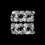
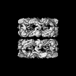
| タイトル | A test-bed for optimizing high-resolution single particle reconstructions. | |||||||||
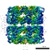
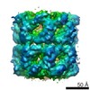
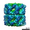
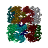
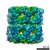
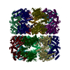
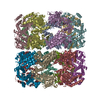
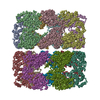
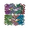
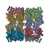
 マップデータ マップデータ | This is a single particle reconstruction of GroEL | |||||||||
 試料 試料 |
| |||||||||
| 生物種 |   Escherichia coli (大腸菌) Escherichia coli (大腸菌) | |||||||||
| 手法 | 単粒子再構成法 / クライオ電子顕微鏡法 / ネガティブ染色法 / 解像度: 5.4 Å | |||||||||
 データ登録者 データ登録者 | Stagg SM / Lander GC / Quispe J / Voss NR / Cheng A / Bradlow H / Bradlow S / Carragher B / Potter CS | |||||||||
 引用 引用 | ジャーナル: J Struct Biol / 年: 2008 タイトル: A test-bed for optimizing high-resolution single particle reconstructions. 著者: Scott M Stagg / Gabriel C Lander / Joel Quispe / Neil R Voss / Anchi Cheng / Henry Bradlow / Steven Bradlow / Bridget Carragher / Clinton S Potter /  要旨: It is becoming routine for cryoEM single particle reconstructions to result in 3D electron density maps with resolutions of approximately 10A, but maps with resolutions of 5A or better are still ...It is becoming routine for cryoEM single particle reconstructions to result in 3D electron density maps with resolutions of approximately 10A, but maps with resolutions of 5A or better are still celebrated events. The electron microscope has a resolving power to better than 2A, and thus should not be a limiting factor; instead the practical limitations in resolution most likely arise from a combination of specimen preparation methods, data collection parameters, and data analysis procedures. With the aid of a highly automated system for acquiring images, coupled to a relational database to keep track of all processing parameters, we have taken a systematic approach to optimizing parameters affecting the resolution of single particle reconstructions. Using GroEL as a test-bed, we performed a series of 3D reconstructions where we systematically varied the number of particles used in computing the map, the accelerating voltage of the microscope, and the electron dose used to acquire the images. We also investigated methods for excluding unacceptable or "bad" particles from contributing to the final 3D map. Using relatively standard instrumentation (Tecnai F20, 4K x 4K CCD, side entry cold stage) and a completely automated approach, these approaches resulted in a map with a nominal resolution of 5.4A (FSC(0.5)) in which secondary structure is clearly discernable and the handedness of some of the alpha-helices in the GroEL structure can be determined. | |||||||||
| 履歴 |
|
- 構造の表示
構造の表示
| ムービー |
 ムービービューア ムービービューア |
|---|---|
| 構造ビューア | EMマップ: SurfViewMolmilJmol/JSmol |
| 添付画像 |
- ダウンロードとリンク
-EMDBアーカイブ
| マップデータ | emd_1457.map.gz | 5.2 MB | EMDBマップデータ形式 | |
|---|---|---|---|---|
| ヘッダ (付随情報) | emd-1457-v30.xmlemd-1457.xml | 9.7 KB 9.7 KB | 表示 表示 | EMDBヘッダ |
| 画像 |  1457.gif 1457.gif | 59.3 KB | ||
| アーカイブディレクトリ |  http://ftp.pdbj.org/pub/emdb/structures/EMD-1457ftp://ftp.pdbj.org/pub/emdb/structures/EMD-1457 http://ftp.pdbj.org/pub/emdb/structures/EMD-1457ftp://ftp.pdbj.org/pub/emdb/structures/EMD-1457 | HTTPS FTP |
-関連構造データ
-リンク
| EMDBのページ | EMDB (EBI/PDBe) / EMDataResource |
|---|
-マップ
| ファイル | ダウンロード / ファイル: emd_1457.map.gz / 形式: CCP4 / 大きさ: 14.1 MB / タイプ: IMAGE STORED AS FLOATING POINT NUMBER (4 BYTES) | ||||||||||||||||||||||||||||||||||||||||||||||||||||||||||||||||||||
|---|---|---|---|---|---|---|---|---|---|---|---|---|---|---|---|---|---|---|---|---|---|---|---|---|---|---|---|---|---|---|---|---|---|---|---|---|---|---|---|---|---|---|---|---|---|---|---|---|---|---|---|---|---|---|---|---|---|---|---|---|---|---|---|---|---|---|---|---|---|
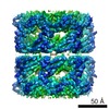
| 注釈 | This is a single particle reconstruction of GroEL | ||||||||||||||||||||||||||||||||||||||||||||||||||||||||||||||||||||
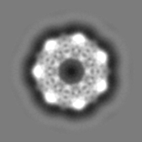
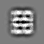
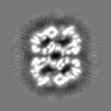
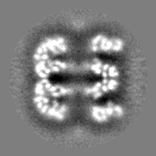
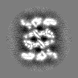
| 投影像・断面図 | 画像のコントロール
画像は Spider により作成 | ||||||||||||||||||||||||||||||||||||||||||||||||||||||||||||||||||||
| ボクセルのサイズ | X=Y=Z: 1.63 Å | ||||||||||||||||||||||||||||||||||||||||||||||||||||||||||||||||||||
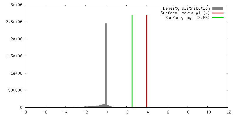
| 密度 |
| ||||||||||||||||||||||||||||||||||||||||||||||||||||||||||||||||||||
| 対称性 | 空間群: 1 | ||||||||||||||||||||||||||||||||||||||||||||||||||||||||||||||||||||
| 詳細 | EMDB XML:
CCP4マップ ヘッダ情報:
| ||||||||||||||||||||||||||||||||||||||||||||||||||||||||||||||||||||
 Z (Sec.)
 Y (Row.)
 X (Col.)


-添付データ
- 試料の構成要素
-全体 : GroEL
| 全体 | 名称: GroEL |
|---|---|
| 要素 |
|
-超分子 #1000: GroEL
| 超分子 | 名称: GroEL / タイプ: sample / ID: 1000 / 集合状態: D7 14-mer / Number unique components: 1 |
|---|---|
| 分子量 | 実験値: 800 KDa / 理論値: 800 KDa |
-分子 #1: GroEL
| 分子 | 名称: GroEL / タイプ: protein_or_peptide / ID: 1 / Name.synonym: GroEL / コピー数: 14 / 集合状態: homotetradecamer / 組換発現: Yes |
|---|---|
| 由来(天然) | 生物種: Escherichia coli (大腸菌) / 細胞: E. coli / 細胞中の位置: cytosol |
| 分子量 | 実験値: 800 KDa / 理論値: 800 KDa |
-実験情報
-構造解析
| 手法 | ネガティブ染色法, クライオ電子顕微鏡法 |
|---|---|
 解析 解析 | 単粒子再構成法 |
| 試料の集合状態 | particle |
-試料調製
| 濃度 | 4 mg/mL |
|---|---|
| 緩衝液 | pH: 7.5 / 詳細: 100mM Hepes, 10mM Mg(OAc)2, 10mM KOAc, 2mM DTT |
| 染色 | タイプ: NEGATIVE / 詳細: not stained |
| グリッド | 詳細: Protochips C-flat grid: holey carbon with 2um holes and 2um spacing 400 mesh copper grid |
| 凍結 | 凍結剤: ETHANE / チャンバー内湿度: 100 % / チャンバー内温度: 93 K / 装置: OTHER 詳細: Vitrification instrument: Vitrobot. Grid plasma cleaned for 20s with Fischione 1020 plasma cleaner using 75% Argon 25% Oxygen mix. 手法: Temperature of chamber was 4 degrees C. 0 seconds drain time. Single blot. 0 mm offset. 4 ul sample applied to grid. Blot for 3.5 seconds before plunging. |
- 電子顕微鏡法
| 顕微鏡 | FEI TECNAI F20 |
|---|---|
| 電子線 | 加速電圧: 120 kV / 電子線源: FIELD EMISSION GUN |
| 電子光学系 | 倍率(補正後): 100000 / 照射モード: FLOOD BEAM / 撮影モード: BRIGHT FIELDBright-field microscopy / Cs: 2 mm / 最大 デフォーカス(公称値): 3.0 µm / 最小 デフォーカス(公称値): 1.0 µm / 倍率(公称値): 100000 |
| 試料ステージ | 試料ホルダー: Side entry liquid nitrogen-cooled cryo specimen holder 試料ホルダーモデル: GATAN LIQUID NITROGEN |
| 温度 | 平均: 102 K |
| アライメント法 | Legacy - 非点収差: objective lens astigmatism was corrected automatically using Leginon |
| 日付 | 2006年7月12日 |
| 撮影 | カテゴリ: CCD フィルム・検出器のモデル: GATAN ULTRASCAN 4000 (4k x 4k) 平均電子線量: 13 e/Å2 |
| Tilt angle min | 0 |
| Tilt angle max | 0 |
| 実験機器 |  モデル: Tecnai F20 / 画像提供: FEI Company |
-画像解析
| CTF補正 | 詳細: Phase correction for each particle. |
|---|---|
| 最終 2次元分類 | クラス数: 1294 |
| 最終 再構成 | 想定した対称性 - 点群: D7 (2回x7回 2面回転対称) アルゴリズム: OTHER / 解像度のタイプ: BY AUTHOR / 解像度: 5.4 Å / 解像度の算出法: FSC 0.5 CUT-OFF / ソフトウェア - 名称: EMAN / 使用した粒子像数: 55351 |
