National Health and Medical Research Council (NHMRC, Australia)
APP1128120
Australia
National Health and Medical Research Council (NHMRC, Australia)
APP1146578
Australia
Citation
Journal: Nat Struct Mol Biol / Year: 2022 Title: Structure of the metastatic factor P-Rex1 reveals a two-layered autoinhibitory mechanism. Authors: Yong-Gang Chang / Christopher J Lupton / Charles Bayly-Jones / Alastair C Keen / Laura D'Andrea / Christina M Lucato / Joel R Steele / Hari Venugopal / Ralf B Schittenhelm / James C ...Authors: Yong-Gang Chang / Christopher J Lupton / Charles Bayly-Jones / Alastair C Keen / Laura D'Andrea / Christina M Lucato / Joel R Steele / Hari Venugopal / Ralf B Schittenhelm / James C Whisstock / Michelle L Halls / Andrew M Ellisdon / Abstract: P-Rex (PI(3,4,5)P-dependent Rac exchanger) guanine nucleotide exchange factors potently activate Rho GTPases. P-Rex guanine nucleotide exchange factors are autoinhibited, synergistically activated by ...P-Rex (PI(3,4,5)P-dependent Rac exchanger) guanine nucleotide exchange factors potently activate Rho GTPases. P-Rex guanine nucleotide exchange factors are autoinhibited, synergistically activated by Gβγ and PI(3,4,5)P binding and dysregulated in cancer. Here, we use X-ray crystallography, cryogenic electron microscopy and crosslinking mass spectrometry to determine the structural basis of human P-Rex1 autoinhibition. P-Rex1 has a bipartite structure of N- and C-terminal modules connected by a C-terminal four-helix bundle that binds the N-terminal Pleckstrin homology (PH) domain. In the N-terminal module, the Dbl homology (DH) domain catalytic surface is occluded by the compact arrangement of the DH-PH-DEP1 domains. Structural analysis reveals a remarkable conformational transition to release autoinhibition, requiring a 126° opening of the DH domain hinge helix. The off-axis position of Gβγ and PI(3,4,5)P binding sites further suggests a counter-rotation of the P-Rex1 halves by 90° facilitates PH domain uncoupling from the four-helix bundle, releasing the autoinhibited DH domain to drive Rho GTPase signaling.
Mass: 96.063 Da / Num. of mol.: 6 / Source method: obtained synthetically / Formula: SO4
Has ligand of interest
N
Sequence details
T4-Lysozyme is spliced/inserted into the P-Rex1 structure in a loop region to enable ...T4-Lysozyme is spliced/inserted into the P-Rex1 structure in a loop region to enable crystallisation. However, the T4L could not be built into the PDB model as it was too flexible. Density was present but too poor to build/model the T4L.
-
Experimental details
-
Experiment
Experiment
Method: X-RAY DIFFRACTION / Number of used crystals: 1
-
Sample preparation
Crystal
Density Matthews: 3.86 Å3/Da / Density % sol: 68.1 %
Crystal grow
Temperature: 293 K / Method: vapor diffusion, hanging drop / Details: 1.8 M (NH4)2SO4, 0.05 MES pH 6.0
-
Data collection
Diffraction
Mean temperature: 100 K / Serial crystal experiment: N
Diffraction source
Source: SYNCHROTRON / Site: Australian Synchrotron / Beamline: MX2 / Wavelength: 0.95373 Å
In the structure databanks used in Yorodumi, some data are registered as the other names, "COVID-19 virus" and "2019-nCoV". Here are the details of the virus and the list of structure data.
Jan 31, 2019. EMDB accession codes are about to change! (news from PDBe EMDB page)
EMDB accession codes are about to change! (news from PDBe EMDB page)
The allocation of 4 digits for EMDB accession codes will soon come to an end. Whilst these codes will remain in use, new EMDB accession codes will include an additional digit and will expand incrementally as the available range of codes is exhausted. The current 4-digit format prefixed with “EMD-” (i.e. EMD-XXXX) will advance to a 5-digit format (i.e. EMD-XXXXX), and so on. It is currently estimated that the 4-digit codes will be depleted around Spring 2019, at which point the 5-digit format will come into force.
The EM Navigator/Yorodumi systems omit the EMD- prefix.
Related info.:Q: What is EMD? / ID/Accession-code notation in Yorodumi/EM Navigator
Yorodumi is a browser for structure data from EMDB, PDB, SASBDB, etc.
This page is also the successor to EM Navigator detail page, and also detail information page/front-end page for Omokage search.
The word "yorodu" (or yorozu) is an old Japanese word meaning "ten thousand". "mi" (miru) is to see.
Related info.:EMDB / PDB / SASBDB / Comparison of 3 databanks / Yorodumi Search / Aug 31, 2016. New EM Navigator & Yorodumi / Yorodumi Papers / Jmol/JSmol / Function and homology information / Changes in new EM Navigator and Yorodumi
 Movie
Movie Controller


 Open data
 Basic information
 Components
 Keywords
 SIGNALING PROTEIN / P-Rex1 / P-Rex2 / GEF /
  Function and homology information

 Authors
 Australia, 2items
  Citation
 Structure visualization
 Downloads & links
 Other downloads
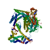

 PDBj
PDBj


 Assembly
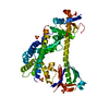


 Mass: 96.063 Da / Num. of mol.: 6 / Source method: obtained synthetically / Formula: SO4
 Sample preparation
 Processing