 Movie
Movie Controller

[English] 日本語
 Yorodumi
- PDB-7syf: Reconstruction of full-length Prex-1 (PtdIns(3,4,5)P3-dependent R... -
+ Open data

- Basic information
| Entry | Database: PDB / ID: 7syf | |||||||||
|---|---|---|---|---|---|---|---|---|---|---|
| Title | Reconstruction of full-length Prex-1 (PtdIns(3,4,5)P3-dependent Rac Exchanger 1) | |||||||||
 Components Components | Phosphatidylinositol 3,4,5-trisphosphate-dependent Rac exchanger 1 protein,Endolysin chimera | |||||||||
 Keywords Keywords | ONCOPROTEIN / guanine nucleotide exchange factor / metastasis / plasma membrane / Rho GTPase signalling | |||||||||
| Function / homology |  Function and homology information Function and homology informationregulation of dendrite development / neutrophil activation / regulation of actin filament polymerization / negative regulation of TOR signaling / regulation of small GTPase mediated signal transduction / superoxide metabolic process / RHOB GTPase cycle / NRAGE signals death through JNK / RHOC GTPase cycle / RHOJ GTPase cycle ...regulation of dendrite development / neutrophil activation / regulation of actin filament polymerization / negative regulation of TOR signaling / regulation of small GTPase mediated signal transduction / superoxide metabolic process / RHOB GTPase cycle / NRAGE signals death through JNK / RHOC GTPase cycle / RHOJ GTPase cycle / RHOQ GTPase cycle / CDC42 GTPase cycle / RHOG GTPase cycle / RAC3 GTPase cycle / RHOA GTPase cycle / RAC2 GTPase cycle / protein serine/threonine kinase inhibitor activity / viral release from host cell by cytolysis / peptidoglycan catabolic process / actin filament polymerization / RAC1 GTPase cycle / guanyl-nucleotide exchange factor activity / GTPase activator activity / dendritic shaft / phospholipid binding / cell wall macromolecule catabolic process / lysozyme / lysozyme activity / G alpha (12/13) signalling events / growth cone / host cell cytoplasm / defense response to bacterium / intracellular signal transduction / positive regulation of cell migration / G protein-coupled receptor signaling pathway / perinuclear region of cytoplasm / enzyme binding / plasma membrane / cytosol Similarity search - Function | |||||||||
| Biological species |  Homo sapiens (human) Homo sapiens (human) | |||||||||
| Method | ELECTRON MICROSCOPY / single particle reconstruction / cryo EM / Resolution: 4.2 Å | |||||||||
 Authors Authors | Lupton, C.J. / Bayly-Jones, C. / Ellisdon, A.M. | |||||||||
| Funding support |  Australia, 2items Australia, 2items
| |||||||||
 Citation Citation | Journal: Nat Struct Mol Biol / Year: 2022 Title: Structure of the metastatic factor P-Rex1 reveals a two-layered autoinhibitory mechanism. Authors: Yong-Gang Chang / Christopher J Lupton / Charles Bayly-Jones / Alastair C Keen / Laura D'Andrea / Christina M Lucato / Joel R Steele / Hari Venugopal / Ralf B Schittenhelm / James C ...Authors: Yong-Gang Chang / Christopher J Lupton / Charles Bayly-Jones / Alastair C Keen / Laura D'Andrea / Christina M Lucato / Joel R Steele / Hari Venugopal / Ralf B Schittenhelm / James C Whisstock / Michelle L Halls / Andrew M Ellisdon / Abstract: P-Rex (PI(3,4,5)P-dependent Rac exchanger) guanine nucleotide exchange factors potently activate Rho GTPases. P-Rex guanine nucleotide exchange factors are autoinhibited, synergistically activated by ...P-Rex (PI(3,4,5)P-dependent Rac exchanger) guanine nucleotide exchange factors potently activate Rho GTPases. P-Rex guanine nucleotide exchange factors are autoinhibited, synergistically activated by Gβγ and PI(3,4,5)P binding and dysregulated in cancer. Here, we use X-ray crystallography, cryogenic electron microscopy and crosslinking mass spectrometry to determine the structural basis of human P-Rex1 autoinhibition. P-Rex1 has a bipartite structure of N- and C-terminal modules connected by a C-terminal four-helix bundle that binds the N-terminal Pleckstrin homology (PH) domain. In the N-terminal module, the Dbl homology (DH) domain catalytic surface is occluded by the compact arrangement of the DH-PH-DEP1 domains. Structural analysis reveals a remarkable conformational transition to release autoinhibition, requiring a 126° opening of the DH domain hinge helix. The off-axis position of Gβγ and PI(3,4,5)P binding sites further suggests a counter-rotation of the P-Rex1 halves by 90° facilitates PH domain uncoupling from the four-helix bundle, releasing the autoinhibited DH domain to drive Rho GTPase signaling. | |||||||||
| History |
|
- Structure visualization
| Structure viewer | Molecule: MolmilJmol/JSmol |
|---|
- Downloads & links
-Download
| PDBx/mmCIF format | 7syf.cif.gz | 264.5 KB | Display | PDBx/mmCIF format |
|---|---|---|---|---|
| PDB format | pdb7syf.ent.gz | 203.7 KB | Display | PDB format |
| PDBx/mmJSON format | 7syf.json.gz | Tree view | PDBx/mmJSON format | |
| Others |  Other downloads Other downloads |
-Validation report
| Arichive directory | https://data.pdbj.org/pub/pdb/validation_reports/sy/7syfftp://data.pdbj.org/pub/pdb/validation_reports/sy/7syf | HTTPS FTP |
|---|
-Related structure data
| Related structure data |  25524MC 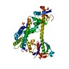 7rx9C M: map data used to model this data C: citing same article ( |
|---|---|
| Similar structure data |


-Links
 PDBj
PDBj


- Assembly
| Deposited unit | 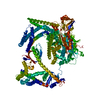
|
|---|---|
| 1 |
|
-Components
| #1: Protein | Mass: 191462.984 Da / Num. of mol.: 1 Source method: isolated from a genetically manipulated source Source: (gene. exp.) Homo sapiens (human) / Gene: PREX1, KIAA1415, e, T4Tp126 / Production host:   Spodoptera frugiperda (fall armyworm) / References: UniProt: Q8TCU6, UniProt: D9IEF7, lysozyme Spodoptera frugiperda (fall armyworm) / References: UniProt: Q8TCU6, UniProt: D9IEF7, lysozyme |
|---|
-Experimental details
-Experiment
| Experiment | Method: ELECTRON MICROSCOPY |
|---|---|
| EM experiment | Aggregation state: PARTICLE / 3D reconstruction method: single particle reconstruction |
- Sample preparation
| Component | Name: full-length Prex-1 (PtdIns(3,4,5)P3-dependent Rac Exchanger 1) Type: COMPLEX / Entity ID: all / Source: RECOMBINANT |
|---|---|
| Molecular weight | Value: 0.191 MDa / Experimental value: YES |
| Source (natural) | Organism: Homo sapiens (human) |
| Source (recombinant) | Organism: Spodoptera frugiperda (fall armyworm) |
| Buffer solution | pH: 8 |
| Specimen | Conc.: 0.18 mg/ml / Embedding applied: NO / Shadowing applied: NO / Staining applied: NO / Vitrification applied: YES |
| Vitrification | Cryogen name: ETHANE |
- Electron microscopy imaging
| Experimental equipment |  Model: Titan Krios / Image courtesy: FEI Company |
|---|---|
| Microscopy | Model: FEI TITAN KRIOS |
| Electron gun | Electron source:  FIELD EMISSION GUN / Accelerating voltage: 300 kV / Illumination mode: FLOOD BEAM FIELD EMISSION GUN / Accelerating voltage: 300 kV / Illumination mode: FLOOD BEAM |
| Electron lens | Mode: BRIGHT FIELD |
| Image recording | Electron dose: 50 e/Å2 / Film or detector model: GATAN K3 BIOQUANTUM (6k x 4k) |
- Processing
| CTF correction | Type: PHASE FLIPPING AND AMPLITUDE CORRECTION |
|---|---|
| Symmetry | Point symmetry: C1 (asymmetric) |
| 3D reconstruction | Resolution: 4.2 Å / Resolution method: FSC 0.143 CUT-OFF / Num. of particles: 123896 / Symmetry type: POINT |
