+ Open data
- Basic information
| Entry | Database: PDB / ID: 7rx9 | |||||||||
|---|---|---|---|---|---|---|---|---|---|---|
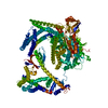
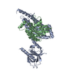
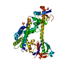
| Title | Structure of autoinhibited P-Rex1 | |||||||||
 Components Components | Phosphatidylinositol 3,4,5-trisphosphate-dependent Rac exchanger 1 protein, Endolysin chimera | |||||||||
 Keywords Keywords | SIGNALING PROTEIN / P-Rex1 / P-Rex2 / GEF / cell growth / Rac1 / Cdc42 | |||||||||
| Function / homology |  Function and homology information Function and homology informationregulation of dendrite development / regulation of actin filament polymerization / neutrophil activation / regulation of small GTPase mediated signal transduction / negative regulation of TOR signaling / RHOB GTPase cycle / superoxide metabolic process / NRAGE signals death through JNK / RHOC GTPase cycle / RHOJ GTPase cycle ...regulation of dendrite development / regulation of actin filament polymerization / neutrophil activation / regulation of small GTPase mediated signal transduction / negative regulation of TOR signaling / RHOB GTPase cycle / superoxide metabolic process / NRAGE signals death through JNK / RHOC GTPase cycle / RHOJ GTPase cycle / RHOQ GTPase cycle / CDC42 GTPase cycle / T cell differentiation / RHOG GTPase cycle / RHOA GTPase cycle / RAC2 GTPase cycle / RAC3 GTPase cycle / protein serine/threonine kinase inhibitor activity / viral release from host cell by cytolysis / peptidoglycan catabolic process / actin filament polymerization / neutrophil chemotaxis / RAC1 GTPase cycle / positive regulation of substrate adhesion-dependent cell spreading / GTPase activator activity / guanyl-nucleotide exchange factor activity / dendritic shaft / phospholipid binding / cell wall macromolecule catabolic process / lysozyme / lysozyme activity / G alpha (12/13) signalling events / growth cone / host cell cytoplasm / defense response to bacterium / intracellular signal transduction / positive regulation of cell migration / G protein-coupled receptor signaling pathway / perinuclear region of cytoplasm / enzyme binding / plasma membrane / cytosol Similarity search - Function | |||||||||
| Biological species |  Homo sapiens (human) Homo sapiens (human) Enterobacteria phage T4 (virus) Enterobacteria phage T4 (virus) | |||||||||
| Method |  X-RAY DIFFRACTION / X-RAY DIFFRACTION /  SYNCHROTRON / SYNCHROTRON /  MOLECULAR REPLACEMENT / Resolution: 3.22 Å MOLECULAR REPLACEMENT / Resolution: 3.22 Å | |||||||||
 Authors Authors | Ellisdon, A.M. / Chang, Y. | |||||||||
| Funding support |  Australia, 2items Australia, 2items
| |||||||||
 Citation Citation |  Journal: Nat Struct Mol Biol / Year: 2022 Journal: Nat Struct Mol Biol / Year: 2022Title: Structure of the metastatic factor P-Rex1 reveals a two-layered autoinhibitory mechanism. Authors: Yong-Gang Chang / Christopher J Lupton / Charles Bayly-Jones / Alastair C Keen / Laura D'Andrea / Christina M Lucato / Joel R Steele / Hari Venugopal / Ralf B Schittenhelm / James C ...Authors: Yong-Gang Chang / Christopher J Lupton / Charles Bayly-Jones / Alastair C Keen / Laura D'Andrea / Christina M Lucato / Joel R Steele / Hari Venugopal / Ralf B Schittenhelm / James C Whisstock / Michelle L Halls / Andrew M Ellisdon /  Abstract: P-Rex (PI(3,4,5)P-dependent Rac exchanger) guanine nucleotide exchange factors potently activate Rho GTPases. P-Rex guanine nucleotide exchange factors are autoinhibited, synergistically activated by ...P-Rex (PI(3,4,5)P-dependent Rac exchanger) guanine nucleotide exchange factors potently activate Rho GTPases. P-Rex guanine nucleotide exchange factors are autoinhibited, synergistically activated by Gβγ and PI(3,4,5)P binding and dysregulated in cancer. Here, we use X-ray crystallography, cryogenic electron microscopy and crosslinking mass spectrometry to determine the structural basis of human P-Rex1 autoinhibition. P-Rex1 has a bipartite structure of N- and C-terminal modules connected by a C-terminal four-helix bundle that binds the N-terminal Pleckstrin homology (PH) domain. In the N-terminal module, the Dbl homology (DH) domain catalytic surface is occluded by the compact arrangement of the DH-PH-DEP1 domains. Structural analysis reveals a remarkable conformational transition to release autoinhibition, requiring a 126° opening of the DH domain hinge helix. The off-axis position of Gβγ and PI(3,4,5)P binding sites further suggests a counter-rotation of the P-Rex1 halves by 90° facilitates PH domain uncoupling from the four-helix bundle, releasing the autoinhibited DH domain to drive Rho GTPase signaling. | |||||||||
| History |
|
- Structure visualization
| Structure viewer | Molecule:  Molmil Molmil Jmol/JSmol Jmol/JSmol |
|---|
- Downloads & links
- Download
| PDBx/mmCIF format |  7rx9.cif.gz 7rx9.cif.gz | 221.6 KB | Display |  PDBx/mmCIF format PDBx/mmCIF format |
|---|---|---|---|---|
| PDB format |  pdb7rx9.ent.gz pdb7rx9.ent.gz | 157.3 KB | Display |  PDB format PDB format |
| PDBx/mmJSON format |  7rx9.json.gz 7rx9.json.gz | Tree view |  PDBx/mmJSON format PDBx/mmJSON format | |
| Others |  Other downloads Other downloads |
-Validation report
| Arichive directory |  https://data.pdbj.org/pub/pdb/validation_reports/rx/7rx9 https://data.pdbj.org/pub/pdb/validation_reports/rx/7rx9 ftp://data.pdbj.org/pub/pdb/validation_reports/rx/7rx9 ftp://data.pdbj.org/pub/pdb/validation_reports/rx/7rx9 | HTTPS FTP |
|---|
-Related structure data
| Related structure data |  7syfC  4yonS S: Starting model for refinement C: citing same article ( |
|---|---|
| Similar structure data | Similarity search - Function & homology  F&H Search F&H Search |
- Links
Links
- Assembly
| Deposited unit | 
| ||||||||||||
|---|---|---|---|---|---|---|---|---|---|---|---|---|---|
| 1 |
| ||||||||||||
| Unit cell |
|
- Components
| #1: Protein | Mass: 70026.695 Da / Num. of mol.: 1 Source method: isolated from a genetically manipulated source Source: (gene. exp.)  Homo sapiens (human), (gene. exp.) Homo sapiens (human), (gene. exp.)  Enterobacteria phage T4 (virus) Enterobacteria phage T4 (virus)Gene: PREX1, KIAA1415 / Production host:  | ||||
|---|---|---|---|---|---|
| #2: Chemical | ChemComp-SO4 / Has ligand of interest | N | Sequence details | T4-Lysozyme is spliced/inserted into the P-Rex1 structure in a loop region to enable ...T4-Lysozyme is spliced/inserted into the P-Rex1 structure in a loop region to enable crystallisation. However, the T4L could not be built into the PDB model as it was too flexible. Density was present but too poor to build/model the T4L. | |
-Experimental details
-Experiment
| Experiment | Method:  X-RAY DIFFRACTION / Number of used crystals: 1 X-RAY DIFFRACTION / Number of used crystals: 1 |
|---|
- Sample preparation
| Crystal | Density Matthews: 3.86 Å3/Da / Density % sol: 68.1 % |
|---|---|
| Crystal grow | Temperature: 293 K / Method: vapor diffusion, hanging drop / Details: 1.8 M (NH4)2SO4, 0.05 MES pH 6.0 |
-Data collection
| Diffraction | Mean temperature: 100 K / Serial crystal experiment: N |
|---|---|
| Diffraction source | Source:  SYNCHROTRON / Site: SYNCHROTRON / Site:  Australian Synchrotron Australian Synchrotron  / Beamline: MX2 / Wavelength: 0.95373 Å / Beamline: MX2 / Wavelength: 0.95373 Å |
| Detector | Type: DECTRIS EIGER X 16M / Detector: PIXEL / Date: Jul 7, 2021 |
| Radiation | Protocol: SINGLE WAVELENGTH / Monochromatic (M) / Laue (L): M / Scattering type: x-ray |
| Radiation wavelength | Wavelength: 0.95373 Å / Relative weight: 1 |
| Reflection | Resolution: 3.22→47.83 Å / Num. obs: 18039 / % possible obs: 99.1 % / Redundancy: 6.4 % / Biso Wilson estimate: 119.41 Å2 / CC1/2: 0.998 / Net I/σ(I): 7.8 |
| Reflection shell | Resolution: 3.22→3.48 Å / Num. unique obs: 3597 / CC1/2: 0.358 |
- Processing
| Software |
| |||||||||||||||||||||||||||||||||||||||||||||||||||||||||||||||||||||||||||
|---|---|---|---|---|---|---|---|---|---|---|---|---|---|---|---|---|---|---|---|---|---|---|---|---|---|---|---|---|---|---|---|---|---|---|---|---|---|---|---|---|---|---|---|---|---|---|---|---|---|---|---|---|---|---|---|---|---|---|---|---|---|---|---|---|---|---|---|---|---|---|---|---|---|---|---|---|
| Refinement | Method to determine structure:  MOLECULAR REPLACEMENT MOLECULAR REPLACEMENTStarting model: 4YON Resolution: 3.22→47.83 Å / SU ML: 0.4288 / Cross valid method: FREE R-VALUE / σ(F): 1.33 / Phase error: 30.4388 Stereochemistry target values: GeoStd + Monomer Library + CDL v1.2
| |||||||||||||||||||||||||||||||||||||||||||||||||||||||||||||||||||||||||||
| Solvent computation | Shrinkage radii: 0.9 Å / VDW probe radii: 1.11 Å / Solvent model: FLAT BULK SOLVENT MODEL | |||||||||||||||||||||||||||||||||||||||||||||||||||||||||||||||||||||||||||
| Displacement parameters | Biso mean: 127.08 Å2 | |||||||||||||||||||||||||||||||||||||||||||||||||||||||||||||||||||||||||||
| Refinement step | Cycle: LAST / Resolution: 3.22→47.83 Å
| |||||||||||||||||||||||||||||||||||||||||||||||||||||||||||||||||||||||||||
| Refine LS restraints |
| |||||||||||||||||||||||||||||||||||||||||||||||||||||||||||||||||||||||||||
| LS refinement shell |
| |||||||||||||||||||||||||||||||||||||||||||||||||||||||||||||||||||||||||||
| Refinement TLS params. | Method: refined / Refine-ID: X-RAY DIFFRACTION
| |||||||||||||||||||||||||||||||||||||||||||||||||||||||||||||||||||||||||||
| Refinement TLS group | Refine-ID: X-RAY DIFFRACTION / Auth asym-ID: A / Label asym-ID: A
|
 Movie
Movie Controller


 PDBj
PDBj


