 Movie
Movie Controller


+ Open data

- Basic information
| Entry | Database: PDB / ID: 4ug0 | ||||||
|---|---|---|---|---|---|---|---|
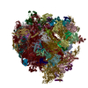
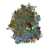
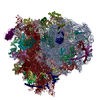
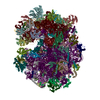
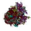
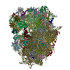
| Title | STRUCTURE OF THE HUMAN 80S RIBOSOME | ||||||
 Components Components |
| ||||||
 Keywords Keywords |  RIBOSOME RIBOSOME | ||||||
| Function / homology |  Function and homology information Function and homology informationpositive regulation of cysteine-type endopeptidase activity involved in execution phase of apoptosis / negative regulation of endoplasmic reticulum unfolded protein response / eukaryotic 80S initiation complex / oxidized pyrimidine DNA binding / response to TNF agonist / positive regulation of base-excision repair / negative regulation of protein neddylation / protein tyrosine kinase inhibitor activity / translation at presynapse / axial mesoderm development ...positive regulation of cysteine-type endopeptidase activity involved in execution phase of apoptosis / negative regulation of endoplasmic reticulum unfolded protein response / eukaryotic 80S initiation complex / oxidized pyrimidine DNA binding / response to TNF agonist / positive regulation of base-excision repair / negative regulation of protein neddylation / protein tyrosine kinase inhibitor activity / translation at presynapse / axial mesoderm development / positive regulation of intrinsic apoptotic signaling pathway in response to DNA damage / positive regulation of respiratory burst involved in inflammatory response / positive regulation of gastrulation / regulation of G1 to G0 transition / negative regulation of formation of translation preinitiation complex / ribosomal protein import into nucleus / positive regulation of intrinsic apoptotic signaling pathway in response to DNA damage by p53 class mediator / regulation of translation involved in cellular response to UV / IRE1-RACK1-PP2A complex / nucleolus organization / : / exit from mitosis / positive regulation of endodeoxyribonuclease activity / positive regulation of Golgi to plasma membrane protein transport / protein-DNA complex disassembly / 90S preribosome assembly / positive regulation of DNA damage response, signal transduction by p53 class mediator resulting in transcription of p21 class mediator / TNFR1-mediated ceramide production / negative regulation of RNA splicing / negative regulation of DNA repair / laminin receptor activity / optic nerve development / SRP-dependent cotranslational protein targeting to membrane / TORC2 complex binding / oxidized purine DNA binding / GAIT complex / G1 to G0 transition / negative regulation of intrinsic apoptotic signaling pathway in response to hydrogen peroxide / supercoiled DNA binding / neural crest cell differentiation / retinal ganglion cell axon guidance / middle ear morphogenesis / negative regulation of phagocytosis / NF-kappaB complex / rRNA modification in the nucleus and cytosol / ubiquitin-like protein conjugating enzyme binding / regulation of establishment of cell polarity / positive regulation of ubiquitin-protein transferase activity / Formation of the ternary complex, and subsequently, the 43S complex / erythrocyte homeostasis / cytoplasmic side of rough endoplasmic reticulum membrane / A band / positive regulation of signal transduction by p53 class mediator / alpha-beta T cell differentiation / ubiquitin ligase inhibitor activity / nuclear-transcribed mRNA catabolic process, nonsense-mediated decay / pigmentation / protein kinase A binding / Ribosomal scanning and start codon recognition / negative regulation of ubiquitin protein ligase activity / ion channel inhibitor activity / Translation initiation complex formation / phagocytic cup / positive regulation of mitochondrial depolarization / response to aldosterone / homeostatic process / negative regulation of Wnt signaling pathway / lung morphogenesis / macrophage chemotaxis / positive regulation of T cell receptor signaling pathway / fibroblast growth factor binding / positive regulation of activated T cell proliferation / regulation of cell division / Protein hydroxylation / male meiosis I / TOR signaling / iron-sulfur cluster binding / BH3 domain binding / mTORC1-mediated signalling / SARS-CoV-1 modulates host translation machinery / Peptide chain elongation / viral transcription / positive regulation of intrinsic apoptotic signaling pathway by p53 class mediator / endonucleolytic cleavage to generate mature 3'-end of SSU-rRNA from (SSU-rRNA, 5.8S rRNA, LSU-rRNA) / Selenocysteine synthesis / protein-RNA complex assembly / monocyte chemotaxis / Formation of a pool of free 40S subunits / cysteine-type endopeptidase activator activity involved in apoptotic process / positive regulation of cyclic-nucleotide phosphodiesterase activity / Eukaryotic Translation Termination / ribosomal small subunit export from nucleus / blastocyst development / Response of EIF2AK4 (GCN2) to amino acid deficiency / SRP-dependent cotranslational protein targeting to membrane / translation regulator activity / Viral mRNA Translation / protein localization to nucleus / cellular response to actinomycin D / Nonsense Mediated Decay (NMD) independent of the Exon Junction Complex (EJC)Similarity search - Function | ||||||
| Biological species |  HOMO SAPIENS (human) HOMO SAPIENS (human) | ||||||
| Method | ELECTRON MICROSCOPY / single particle reconstruction / cryo EM / Resolution: 3.6 Å | ||||||
 Authors Authors | Khatter, H. / Myasnikov, A.G. / Natchiar, S.K. / Klaholz, B.P. | ||||||
 Citation Citation | Journal: Nature / Year: 2015 Title: Structure of the human 80S ribosome. Authors: Heena Khatter / Alexander G Myasnikov / S Kundhavai Natchiar / Bruno P Klaholz /  Abstract: Ribosomes are translational machineries that catalyse protein synthesis. Ribosome structures from various species are known at the atomic level, but obtaining the structure of the human ribosome has ...Ribosomes are translational machineries that catalyse protein synthesis. Ribosome structures from various species are known at the atomic level, but obtaining the structure of the human ribosome has remained a challenge; efforts to address this would be highly relevant with regard to human diseases. Here we report the near-atomic structure of the human ribosome derived from high-resolution single-particle cryo-electron microscopy and atomic model building. The structure has an average resolution of 3.6 Å, reaching 2.9 Å resolution in the most stable regions. It provides unprecedented insights into ribosomal RNA entities and amino acid side chains, notably of the transfer RNA binding sites and specific molecular interactions with the exit site tRNA. It reveals atomic details of the subunit interface, which is seen to remodel strongly upon rotational movements of the ribosomal subunits. Furthermore, the structure paves the way for analysing antibiotic side effects and diseases associated with deregulated protein synthesis. | ||||||
| History |
|
- Structure visualization
| Movie |
Movie viewer |
|---|---|
| Structure viewer | Molecule: MolmilJmol/JSmol |
- Downloads & links
-Download
| PDBx/mmCIF format | 4ug0.cif.gz | 5.3 MB | Display | PDBx/mmCIF format |
|---|---|---|---|---|
| PDB format | pdb4ug0.ent.gz | Display | PDB format | |
| PDBx/mmJSON format | 4ug0.json.gz | Tree view | PDBx/mmJSON format | |
| Others |  Other downloads Other downloads |
-Validation report
| Arichive directory | https://data.pdbj.org/pub/pdb/validation_reports/ug/4ug0ftp://data.pdbj.org/pub/pdb/validation_reports/ug/4ug0 | HTTPS FTP |
|---|
-Related structure data
| Related structure data |  2938MC M: map data used to model this data C: citing same article ( |
|---|---|
| Similar structure data |

-Links
 PDBj
PDBj


- Assembly
| Deposited unit | 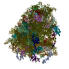
|
|---|---|
| 1 |
|
-Components
-RNA chain , 5 types, 5 molecules L5L7L8S2S6
| #1: RNA chain | / RNA (5070-MER) Mass: 1640238.125 Da / Num. of mol.: 1 / Source method: isolated from a natural source / Source: (natural) HOMO SAPIENS (human) |
|---|---|
| #2: RNA chain | / RNA (121-MER) Mass: 38998.078 Da / Num. of mol.: 1 / Source method: isolated from a natural source / Source: (natural) HOMO SAPIENS (human) |
| #3: RNA chain | / RNA (157-MER) Mass: 50449.812 Da / Num. of mol.: 1 / Source method: isolated from a natural source / Source: (natural) HOMO SAPIENS (human) |
| #47: RNA chain | / RNA (1869-MER) Mass: 602752.875 Da / Num. of mol.: 1 / Source method: isolated from a natural source / Source: (natural) HOMO SAPIENS (human) |
| #48: RNA chain | Mass: 24231.510 Da / Num. of mol.: 1 / Source method: isolated from a natural source / Source: (natural) HOMO SAPIENS (human) / References: GenBank: 174924 |
+60S RIBOSOMAL PROTEIN ... , 42 types, 42 molecules LALBLCLDLELFLGLHLILJLLLMLNLOLPLQLRLSLTLULVLWLXLYLZLaLbLcLdLe...
-Protein , 4 types, 4 molecules LmSfSgSM
| #41: Protein | Mass: 14758.394 Da / Num. of mol.: 1 / Source method: isolated from a natural source / Source: (natural) HOMO SAPIENS (human) / References: UniProt: P62987 |
|---|---|
| #69: Protein | Mass: 18004.041 Da / Num. of mol.: 1 / Source method: isolated from a natural source / Source: (natural) HOMO SAPIENS (human) / References: UniProt: P62979 |
| #70: Protein | Mass: 35115.652 Da / Num. of mol.: 1 / Source method: isolated from a natural source / Source: (natural) HOMO SAPIENS (human) / References: UniProt: P63244 |
| #74: Protein | Mass: 14548.896 Da / Num. of mol.: 1 / Source method: isolated from a natural source / Source: (natural) HOMO SAPIENS (human) |
+40S RIBOSOMAL PROTEIN ... , 30 types, 30 molecules SASBSDSESFSHSISKSLSPSQSRSSSTSUSVSXSaScSdSCSGSJSNSOSWSYSZSbSe
-Non-polymers , 2 types, 245 molecules 

| #82: Chemical | ChemComp-MG /  Mass: 24.305 Da / Num. of mol.: 239 / Source method: obtained synthetically / Formula: Mg Mass: 24.305 Da / Num. of mol.: 239 / Source method: obtained synthetically / Formula: Mg#83: Chemical | ChemComp-ZN /  Mass: 65.409 Da / Num. of mol.: 6 / Source method: obtained synthetically / Formula: Zn Mass: 65.409 Da / Num. of mol.: 6 / Source method: obtained synthetically / Formula: Zn |
|---|
-Experimental details
-Experiment
| Experiment | Method: ELECTRON MICROSCOPY |
|---|---|
| EM experiment | Aggregation state: PARTICLE / 3D reconstruction method: single particle reconstruction |
- Sample preparation
| Component | Name: HUMAN 80S RIBOSOMEEukaryotic ribosome / Type: RIBOSOME |
|---|---|
| Buffer solution | pH: 7.6 |
| Specimen | Conc.: 0.1 mg/ml / Embedding applied: NO / Shadowing applied: NO / Staining applied: NO / Vitrification applied: YES |
| Vitrification | Instrument: FEI VITROBOT MARK IV / Cryogen name: ETHANE |
- Electron microscopy imaging
| Experimental equipment |  Model: Titan Krios / Image courtesy: FEI Company |
|---|---|
| Microscopy | Model: FEI TITAN KRIOS / Date: Jan 10, 2014 |
| Electron gun | Electron source: FIELD EMISSION GUN / Accelerating voltage: 300 kV / Illumination mode: SPOT SCAN |
| Electron lens | Mode: BRIGHT FIELDBright-field microscopy / Nominal magnification: 59000 X / Nominal defocus max: 4500 nm / Nominal defocus min: 600 nm |
| Image recording | Electron dose: 60 e/Å2 / Film or detector model: FEI FALCON II (4k x 4k) |
- Processing
| Symmetry | Point symmetry: C1 (asymmetric) |
|---|---|
| 3D reconstruction | Resolution: 3.6 Å / Resolution method: FSC 0.143 CUT-OFF / Num. of particles: 24000 / Refinement type: HALF-MAPS REFINED INDEPENDENTLY / Symmetry type: POINT |
| Atomic model building | Protocol: FLEXIBLE FIT / Details: METHOD--FLEXIBLE |
