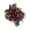
- EMDB-2938: Structure of the human 80S ribosome -
+
Open data
ID or keywords:
Loading...
-
Basic information
Entry
Database: EMDB / ID: EMD-2938
Title
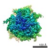
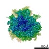
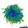
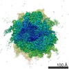
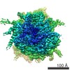
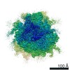
Structure of the human 80S ribosome
Map data
Relion, masked map
Sample
Sample: 80S human
Complex: HeLa Cytoplasmic 80S
Keywords
80S human ribosome / high resolution cryoEM
Function / homology
Function and homology information
translation at presynapse / exit from mitosis / optic nerve development / response to insecticide / regulation of translation involved in cellular response to UV / eukaryotic 80S initiation complex / ribosomal protein import into nucleus / regulation of G1 to G0 transition / negative regulation of endoplasmic reticulum unfolded protein response / negative regulation of formation of translation preinitiation complex ...translation at presynapse / exit from mitosis / optic nerve development / response to insecticide / regulation of translation involved in cellular response to UV / eukaryotic 80S initiation complex / ribosomal protein import into nucleus / regulation of G1 to G0 transition / negative regulation of endoplasmic reticulum unfolded protein response / negative regulation of formation of translation preinitiation complex / axial mesoderm development / retinal ganglion cell axon guidance / oxidized pyrimidine DNA binding / response to TNF agonist / positive regulation of base-excision repair / positive regulation of intrinsic apoptotic signaling pathway in response to DNA damage by p53 class mediator / protein-DNA complex disassembly / positive regulation of respiratory burst involved in inflammatory response / positive regulation of gastrulation / 90S preribosome assembly / protein tyrosine kinase inhibitor activity / positive regulation of intrinsic apoptotic signaling pathway in response to DNA damage / positive regulation of ubiquitin-protein transferase activity / IRE1-RACK1-PP2A complex / positive regulation of DNA-templated transcription initiation / alpha-beta T cell differentiation / positive regulation of Golgi to plasma membrane protein transport / nucleolus organization / TNFR1-mediated ceramide production / positive regulation of DNA damage response, signal transduction by p53 class mediator / GAIT complex / negative regulation of RNA splicing / TORC2 complex binding / neural crest cell differentiation / supercoiled DNA binding / G1 to G0 transition / NF-kappaB complex / negative regulation of DNA repair / cytoplasmic translational initiation / cysteine-type endopeptidase activator activity involved in apoptotic process / oxidized purine DNA binding / middle ear morphogenesis / rRNA modification in the nucleus and cytosol / negative regulation of intrinsic apoptotic signaling pathway in response to hydrogen peroxide / regulation of establishment of cell polarity / negative regulation of bicellular tight junction assembly / ubiquitin-like protein conjugating enzyme binding / negative regulation of phagocytosis / erythrocyte homeostasis / cytoplasmic side of rough endoplasmic reticulum membrane / Formation of the ternary complex, and subsequently, the 43S complex / ion channel inhibitor activity / protein kinase A binding / laminin receptor activity / homeostatic process / pigmentation / Ribosomal scanning and start codon recognition / positive regulation of mitochondrial depolarization / macrophage chemotaxis / Translation initiation complex formation / lung morphogenesis / negative regulation of Wnt signaling pathway / positive regulation of natural killer cell proliferation / male meiosis I / fibroblast growth factor binding / Protein hydroxylation / monocyte chemotaxis / BH3 domain binding / negative regulation of translational frameshifting / regulation of adenylate cyclase-activating G protein-coupled receptor signaling pathway / TOR signaling / mTORC1-mediated signalling / SARS-CoV-1 modulates host translation machinery / iron-sulfur cluster binding / positive regulation of GTPase activity / regulation of cell division / cellular response to ethanol / Peptide chain elongation / Selenocysteine synthesis / Formation of a pool of free 40S subunits / negative regulation of protein binding / blastocyst development / Eukaryotic Translation Termination / positive regulation of intrinsic apoptotic signaling pathway by p53 class mediator / protein serine/threonine kinase inhibitor activity / SRP-dependent cotranslational protein targeting to membrane / Response of EIF2AK4 (GCN2) to amino acid deficiency / negative regulation of respiratory burst involved in inflammatory response / ubiquitin ligase inhibitor activity / Viral mRNA Translation / endonucleolytic cleavage to generate mature 3'-end of SSU-rRNA from (SSU-rRNA, 5.8S rRNA, LSU-rRNA) / protein localization to nucleus / negative regulation of ubiquitin-dependent protein catabolic process / Nonsense Mediated Decay (NMD) independent of the Exon Junction Complex (EJC) / positive regulation of signal transduction by p53 class mediator / GTP hydrolysis and joining of the 60S ribosomal subunit / L13a-mediated translational silencing of Ceruloplasmin expression / Major pathway of rRNA processing in the nucleolus and cytosol / protein targeting / regulation of translational fidelity Similarity search - Function
40S ribosomal protein SA / Ribosomal protein L6, N-terminal / Ribosomal protein L6, N-terminal domain / 40S ribosomal protein SA, C-terminal domain / 40S ribosomal protein SA C-terminus / Ubiquitin-like protein FUBI / Ribosomal protein L30e / Ribosomal L15/L27a, N-terminal / Ribosomal protein L28e / : ...40S ribosomal protein SA / Ribosomal protein L6, N-terminal / Ribosomal protein L6, N-terminal domain / 40S ribosomal protein SA, C-terminal domain / 40S ribosomal protein SA C-terminus / Ubiquitin-like protein FUBI / Ribosomal protein L30e / Ribosomal L15/L27a, N-terminal / Ribosomal protein L28e / : / Ribosomal protein L23 / Ribosomal protein L2, archaeal-type / Ribosomal L28e/Mak16 / Ribosomal L28e protein family / Ribosomal protein L1, conserved site / Ribosomal protein L1 signature. / Ribosomal protein L1 / metallochaperone-like domain / TRASH domain / Ribosomal protein L1, 3-layer alpha/beta-sandwich / : / Ribosomal protein S26e signature. / Ribosomal protein L41 / Ribosomal protein L41 / Ribosomal protein L13e, conserved site / Ribosomal protein L13e signature. / Ribosomal protein S21e, conserved site / Ribosomal protein S21e signature. / Ribosomal protein S26e / Ribosomal protein L29e / Ribosomal protein S26e superfamily / Ribosomal protein S26e / Ribosomal L29e protein family / Ribosomal protein L1-like / Ribosomal protein L1/ribosomal biogenesis protein / Ribosomal protein L1p/L10e family / Ribosomal protein L22e / Ribosomal protein L22e superfamily / Ribosomal L22e protein family / Ribosomal protein L27e, conserved site / Ribosomal protein L27e signature. / Small (40S) ribosomal subunit Asc1/RACK1 / Ribosomal protein L13e / Ribosomal protein L13e / Ribosomal protein L38e / Ribosomal protein S5, eukaryotic/archaeal / Ribosomal protein L38e superfamily / Ribosomal L38e protein family / Ribosomal protein S19e, conserved site / : / Ribosomal protein S19e signature. / Ribosomal protein L19, eukaryotic / Ribosomal protein S21e / Ribosomal protein S21e superfamily / Ribosomal protein S21e / Ribosomal protein L6e signature. / 60S ribosomal protein L18a/ L20, eukaryotes / Ribosomal protein S2, eukaryotic / Ribosomal protein L10e, conserved site / Ribosomal protein L10e signature. / Ribosomal protein L19/L19e conserved site / Ribosomal protein L19e signature. / Ribosomal protein L44e signature. / Ribosomal protein L24e, conserved site / Ribosomal protein L24e signature. / Ribosomal protein L18/L18-A/B/e, conserved site / Ribosomal protein L18e signature. / 40S Ribosomal protein S10 / Ribosomal protein L10e / S27a-like superfamily / Ribosomal protein L34e, conserved site / Ribosomal protein L34e signature. / Plectin/S10, N-terminal / Plectin/S10 domain / Ribosomal protein L5 eukaryotic, C-terminal / Ribosomal L18 C-terminal region / Ribosomal protein L23/L25, N-terminal / Ribosomal protein L23, N-terminal domain / 50S ribosomal protein L18Ae/60S ribosomal protein L20 and L18a / : / Ribosomal L40e family / Ribosomal protein L30e signature 1. / Ribosomal protein 50S-L18Ae/60S-L20/60S-L18A / Ribosomal proteins 50S-L18Ae/60S-L20/60S-L18A / Ribosomal protein L36e signature. / Ribosomal protein S10, eukaryotic/archaeal / Ribosomal protein S30 / Ribosomal protein S30 / Ribosomal protein L44e / Ribosomal protein L44 / Ribosomal protein 60S L18 and 50S L18e / Ribosomal protein L35Ae, conserved site / Ribosomal protein L35Ae signature. / Eukaryotic Ribosomal Protein L27, KOW domain / Ribosomal_L40e / Ribosomal protein L40e / Ribosomal protein L40e superfamily / Ribosomal protein S25 / : / S25 ribosomal protein Similarity search - Domain/homology
Small ribosomal subunit protein eS17 / Small ribosomal subunit protein uS2 / Small ribosomal subunit protein eS17 / Small ribosomal subunit protein uS5 / Large ribosomal subunit protein eL33 / Large ribosomal subunit protein uL30 / Large ribosomal subunit protein uL22 / Small ribosomal subunit protein uS3 / Large ribosomal subunit protein eL13 / Large ribosomal subunit protein uL6 ...Small ribosomal subunit protein eS17 / Small ribosomal subunit protein uS2 / Small ribosomal subunit protein eS17 / Small ribosomal subunit protein uS5 / Large ribosomal subunit protein eL33 / Large ribosomal subunit protein uL30 / Large ribosomal subunit protein uL22 / Small ribosomal subunit protein uS3 / Large ribosomal subunit protein eL13 / Large ribosomal subunit protein uL6 / Large ribosomal subunit protein eL22 / Large ribosomal subunit protein uL4 / Small ribosomal subunit protein eS19 / Large ribosomal subunit protein uL3 / Large ribosomal subunit protein uL13 / Small ribosomal subunit protein eS27 / Large ribosomal subunit protein uL29 / Large ribosomal subunit protein uL15 / Large ribosomal subunit protein uL18 / Large ribosomal subunit protein eL21 / Large ribosomal subunit protein eL28 / Small ribosomal subunit protein uS4 / Small ribosomal subunit protein uS7 / Small ribosomal subunit protein eS10 / Large ribosomal subunit protein eL29 / Large ribosomal subunit protein eL34 / Large ribosomal subunit protein eL14 / Small ribosomal subunit protein uS10 / Small ribosomal subunit protein eS1 / Large ribosomal subunit protein uL24 / Large ribosomal subunit protein eL15 / Large ribosomal subunit protein eL27 / Large ribosomal subunit protein eL43 / Large ribosomal subunit protein eL37 / Small ribosomal subunit protein eS7 / Small ribosomal subunit protein eS8 / Small ribosomal subunit protein uS8 / Small ribosomal subunit protein uS9 / Small ribosomal subunit protein uS11 / Small ribosomal subunit protein uS12 / Small ribosomal subunit protein uS13 / Small ribosomal subunit protein uS14 / Small ribosomal subunit protein uS15 / Small ribosomal subunit protein uS17 / Large ribosomal subunit protein eL8 / Small ribosomal subunit protein eS4, X isoform / Large ribosomal subunit protein uL23 / Small ribosomal subunit protein eS6 / Large ribosomal subunit protein uL14 / Small ribosomal subunit protein uS19 / Small ribosomal subunit protein eS24 / Small ribosomal subunit protein eS25 / Small ribosomal subunit protein eS26 / Small ribosomal subunit protein eS28 / Ubiquitin-like FUBI-ribosomal protein eS30 fusion protein / Large ribosomal subunit protein eL30 / Large ribosomal subunit protein eL39 / Large ribosomal subunit protein eL31 / Large ribosomal subunit protein uL1 / Large ribosomal subunit protein eL32 / Large ribosomal subunit protein uL5 / Large ribosomal subunit protein uL2 / Small ribosomal subunit protein eS32 / Ubiquitin-ribosomal protein eS31 fusion protein / Ubiquitin-ribosomal protein eL40 fusion protein / Large ribosomal subunit protein eL38 / Small ribosomal subunit protein eS21 / Small ribosomal subunit protein RACK1 / Large ribosomal subunit protein eL24 / Large ribosomal subunit protein eL42 / Large ribosomal subunit protein eL19 / Large ribosomal subunit protein eL20 / Large ribosomal subunit protein eL6 / Large ribosomal subunit protein eL18 / Ribosomal protein uL16-like / Large ribosomal subunit protein eL36 Similarity search - Component
Biological species
Homo sapiens (human)
Method
single particle reconstruction / cryo EM / Resolution: 3.6 Å
Journal: Nature / Year: 2015 Title: Structure of the human 80S ribosome. Authors: Heena Khatter / Alexander G Myasnikov / S Kundhavai Natchiar / Bruno P Klaholz / Abstract: Ribosomes are translational machineries that catalyse protein synthesis. Ribosome structures from various species are known at the atomic level, but obtaining the structure of the human ribosome has ...Ribosomes are translational machineries that catalyse protein synthesis. Ribosome structures from various species are known at the atomic level, but obtaining the structure of the human ribosome has remained a challenge; efforts to address this would be highly relevant with regard to human diseases. Here we report the near-atomic structure of the human ribosome derived from high-resolution single-particle cryo-electron microscopy and atomic model building. The structure has an average resolution of 3.6 Å, reaching 2.9 Å resolution in the most stable regions. It provides unprecedented insights into ribosomal RNA entities and amino acid side chains, notably of the transfer RNA binding sites and specific molecular interactions with the exit site tRNA. It reveals atomic details of the subunit interface, which is seen to remodel strongly upon rotational movements of the ribosomal subunits. Furthermore, the structure paves the way for analysing antibiotic side effects and diseases associated with deregulated protein synthesis.
History
Deposition
Mar 17, 2015
-
Header (metadata) release
May 6, 2015
-
Map release
May 13, 2015
-
Update
May 13, 2015
-
Current status
May 13, 2015
Processing site: PDBe / Status: Released
-
Structure visualization
Movie
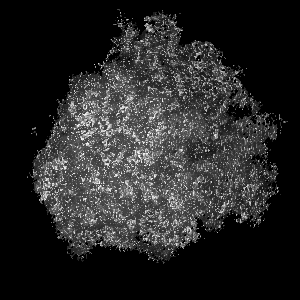
Surface view with section colored by density value
Cryogen name: ETHANE / Chamber humidity: 100 % / Chamber temperature: 120 K / Instrument: FEI VITROBOT MARK IV / Method: Blot Force 5, Blot time - 0.5sec, Wait time 0sec
-
Electron microscopy
Microscope
FEI TITAN KRIOS
Temperature
Min: 80 K / Max: 110 K / Average: 95 K
Alignment procedure
Legacy - Astigmatism: Cs corrector
Date
Jan 10, 2014
Image recording
Category: CCD / Film or detector model: FEI FALCON II (4k x 4k) / Digitization - Sampling interval: 14 µm / Number real images: 3000 / Average electron dose: 60 e/Å2 / Details: 3 frames were collected and total exposure image / Bits/pixel: 16
Electron beam
Acceleration voltage: 300 kV / Electron source: FIELD EMISSION GUN
In the structure databanks used in Yorodumi, some data are registered as the other names, "COVID-19 virus" and "2019-nCoV". Here are the details of the virus and the list of structure data.
Jan 31, 2019. EMDB accession codes are about to change! (news from PDBe EMDB page)
EMDB accession codes are about to change! (news from PDBe EMDB page)
The allocation of 4 digits for EMDB accession codes will soon come to an end. Whilst these codes will remain in use, new EMDB accession codes will include an additional digit and will expand incrementally as the available range of codes is exhausted. The current 4-digit format prefixed with “EMD-” (i.e. EMD-XXXX) will advance to a 5-digit format (i.e. EMD-XXXXX), and so on. It is currently estimated that the 4-digit codes will be depleted around Spring 2019, at which point the 5-digit format will come into force.
The EM Navigator/Yorodumi systems omit the EMD- prefix.
Related info.:Q: What is EMD? / ID/Accession-code notation in Yorodumi/EM Navigator
Yorodumi is a browser for structure data from EMDB, PDB, SASBDB, etc.
This page is also the successor to EM Navigator detail page, and also detail information page/front-end page for Omokage search.
The word "yorodu" (or yorozu) is an old Japanese word meaning "ten thousand". "mi" (miru) is to see.
Related info.:EMDB / PDB / SASBDB / Comparison of 3 databanks / Yorodumi Search / Aug 31, 2016. New EM Navigator & Yorodumi / Yorodumi Papers / Jmol/JSmol / Function and homology information / Changes in new EM Navigator and Yorodumi
 Movie
Movie Controller


 Open data
 Basic information
 Map data
 Sample
 Keywords
 Function and homology information
 Homo sapiens (human)
 Authors
 Citation
 Structure visualization
 Downloads & links
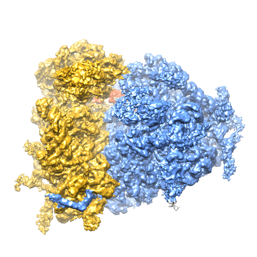 80S_Hum_bp.png
 http://ftp.pdbj.org/pub/emdb/structures/EMD-2938


 Z (Sec.)
 Y (Row.)
 X (Col.)


 Sample components
 Processing
 Electron microscopy
 FIELD EMISSION GUN