Resolution: 2.23→2.27 Å / Redundancy: 2.7 % / Mean I/σ(I) obs: 2.24 / Num. unique all: 1118 / Rsym value: 0.452 / % possible all: 75
-
Processing
Software
Name
Version
Classification
SBC-Collect
datacollection
HKL-3000
datacollection
HKL-3000
phasing
MOLREP
phasing
PHENIX
(phenix.refine: 1.6.4_486)
refinement
HKL-3000
datareduction
HKL-3000
datascaling
Refinement
Method to determine structure: MOLECULAR REPLACEMENT Starting model: The crystal structure of the same protein in the absence of NAD Resolution: 2.227→33.593 Å / SU ML: 0.36 / Isotropic thermal model: mixed / Cross valid method: THROUGHOUT / σ(F): 0 / Stereochemistry target values: ML
Rfactor
Num. reflection
% reflection
Selection details
Rfree
0.254
1490
5.07 %
random
Rwork
0.189
-
-
-
all
0.193
29365
-
-
obs
0.1927
29365
97 %
-
Solvent computation
Shrinkage radii: 0.83 Å / VDW probe radii: 1.1 Å / Solvent model: FLAT BULK SOLVENT MODEL / Bsol: 42.277 Å2 / ksol: 0.318 e/Å3
Displacement parameters
Biso mean: 62.8 Å2
Baniso -1
Baniso -2
Baniso -3
1-
-3.8917 Å2
0 Å2
0.5809 Å2
2-
-
22.5391 Å2
0 Å2
3-
-
-
-18.6474 Å2
Refinement step
Cycle: LAST / Resolution: 2.227→33.593 Å
Protein
Nucleic acid
Ligand
Solvent
Total
Num. atoms
4250
0
94
105
4449
Refine LS restraints
Refine-ID
Type
Dev ideal
Number
X-RAY DIFFRACTION
f_bond_d
0.01
4428
X-RAY DIFFRACTION
f_angle_d
1.249
6007
X-RAY DIFFRACTION
f_dihedral_angle_d
19.725
1756
X-RAY DIFFRACTION
f_chiral_restr
0.074
739
X-RAY DIFFRACTION
f_plane_restr
0.006
747
LS refinement shell
Refine-ID: X-RAY DIFFRACTION
Resolution (Å)
Rfactor Rfree
Num. reflection Rfree
Rfactor Rwork
Num. reflection Rwork
Num. reflection obs
% reflection obs (%)
2.2268-2.3064
0.3424
115
0.26
2190
2305
78
2.3064-2.3987
0.3358
139
0.2585
2692
2831
94
2.3987-2.5078
0.3512
151
0.2493
2867
3018
100
2.5078-2.64
0.3158
124
0.2297
2868
2992
100
2.64-2.8053
0.332
176
0.2284
2816
2992
100
2.8053-3.0218
0.3038
144
0.2199
2848
2992
100
3.0218-3.3257
0.2853
159
0.2259
2859
3018
100
3.3257-3.8063
0.2385
158
0.2029
2903
3061
100
3.8063-4.7933
0.2123
142
0.1509
2905
3047
100
4.7933-33.5969
0.2164
182
0.1516
2927
3109
99
Refinement TLS params.
Method: refined / Refine-ID: X-RAY DIFFRACTION
ID
L11 (°2)
L12 (°2)
L13 (°2)
L22 (°2)
L23 (°2)
L33 (°2)
S11 (Å °)
S12 (Å °)
S13 (Å °)
S21 (Å °)
S22 (Å °)
S23 (Å °)
S31 (Å °)
S32 (Å °)
S33 (Å °)
T11 (Å2)
T12 (Å2)
T13 (Å2)
T22 (Å2)
T23 (Å2)
T33 (Å2)
Origin x (Å)
Origin y (Å)
Origin z (Å)
1
1.7963
-0.0245
0.3093
0.6165
0.0357
1.6554
-0.0783
-0.0137
-0.1196
0.1354
-0.121
-0.0286
-0.0126
-0.0963
0.1354
0.3054
-0.0338
0.0246
0.2213
-0.0484
0.2844
22.7328
-22.9
-24.3181
2
2.2879
0.6052
-0.4739
0.861
-0.6307
1.6351
-0.0974
-0.1344
-0.2605
0.0948
-0.3128
-0.287
-0.1764
0.948
0.3518
0.303
-0.0597
-0.0383
0.5955
0.1984
0.437
54.8644
-25.1214
-27.9546
Refinement TLS group
ID
Refine-ID
Refine TLS-ID
Selection details
1
X-RAY DIFFRACTION
1
chainA
2
X-RAY DIFFRACTION
2
chainB
+
About Yorodumi
-
News
-
Feb 9, 2022. New format data for meta-information of EMDB entries
New format data for meta-information of EMDB entries
Version 3 of the EMDB header file is now the official format.
The previous official version 1.9 will be removed from the archive.
In the structure databanks used in Yorodumi, some data are registered as the other names, "COVID-19 virus" and "2019-nCoV". Here are the details of the virus and the list of structure data.
Jan 31, 2019. EMDB accession codes are about to change! (news from PDBe EMDB page)
EMDB accession codes are about to change! (news from PDBe EMDB page)
The allocation of 4 digits for EMDB accession codes will soon come to an end. Whilst these codes will remain in use, new EMDB accession codes will include an additional digit and will expand incrementally as the available range of codes is exhausted. The current 4-digit format prefixed with “EMD-” (i.e. EMD-XXXX) will advance to a 5-digit format (i.e. EMD-XXXXX), and so on. It is currently estimated that the 4-digit codes will be depleted around Spring 2019, at which point the 5-digit format will come into force.
The EM Navigator/Yorodumi systems omit the EMD- prefix.
Related info.:Q: What is EMD? / ID/Accession-code notation in Yorodumi/EM Navigator
Yorodumi is a browser for structure data from EMDB, PDB, SASBDB, etc.
This page is also the successor to EM Navigator detail page, and also detail information page/front-end page for Omokage search.
The word "yorodu" (or yorozu) is an old Japanese word meaning "ten thousand". "mi" (miru) is to see.
Related info.:EMDB / PDB / SASBDB / Comparison of 3 databanks / Yorodumi Search / Aug 31, 2016. New EM Navigator & Yorodumi / Yorodumi Papers / Jmol/JSmol / Function and homology information / Changes in new EM Navigator and Yorodumi
 Movie
Movie Controller

 Yorodumi
 Open data
 Basic information
 Components
 Keywords
 Function and homology information
 Authors
 Citation
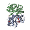
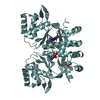
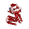
 Structure visualization
 Downloads & links
 Other downloads
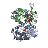
 PDBj
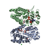
PDBj Assembly
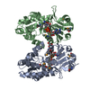

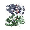
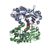
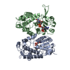
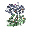
 Mass: 663.425 Da / Num. of mol.: 2 / Source method: obtained synthetically / Formula: C21H27N7O14P2 / Comment: NAD*YM
 Mass: 92.094 Da / Num. of mol.: 1 / Source method: obtained synthetically / Formula: C3H8O3
 Mass: 18.015 Da / Num. of mol.: 105 / Source method: isolated from a natural source / Formula: H2O
 Sample preparation
 / Beamline: 19-ID / Wavelength: 0.97921 Å
 Processing