 Movie
Movie Controller

[English] 日本語
 Yorodumi
- PDB-4b4u: Crystal structure of Acinetobacter baumannii N5, N10- methylenete... -
+ Open data

- Basic information
| Entry | Database: PDB / ID: 4b4u | ||||||
|---|---|---|---|---|---|---|---|
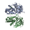
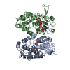
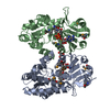
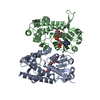
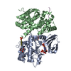
| Title | Crystal structure of Acinetobacter baumannii N5, N10- methylenetetrahydrofolate dehydrogenase-cyclohydrolase (FolD) complexed with NADP cofactor | ||||||
 Components Components | BIFUNCTIONAL PROTEIN FOLD | ||||||
 Keywords Keywords | OXIDOREDUCTASE | ||||||
| Function / homology |  Function and homology information Function and homology informationmethylenetetrahydrofolate dehydrogenase (NADP+) / methenyltetrahydrofolate cyclohydrolase / methenyltetrahydrofolate cyclohydrolase activity / methylenetetrahydrofolate dehydrogenase (NADP+) activity / L-histidine biosynthetic process / : / purine nucleotide biosynthetic process / tetrahydrofolate interconversion / nucleotide binding / cytosol Similarity search - Function | ||||||
| Biological species |  ACINETOBACTER BAUMANNII ATCC 19606 (bacteria) ACINETOBACTER BAUMANNII ATCC 19606 (bacteria) | ||||||
| Method |  X-RAY DIFFRACTION / SYNCHROTRON / MOLECULAR REPLACEMENT / Resolution: 1.45 Å X-RAY DIFFRACTION / SYNCHROTRON / MOLECULAR REPLACEMENT / Resolution: 1.45 Å | ||||||
 Authors Authors | Eadsforth, T.C. / Maluf, F.V. / Hunter, W.N. | ||||||
 Citation Citation | Journal: FEBS J. / Year: 2012 Title: Acinetobacter Baumannii Fold Ligand Complexes; Potent Inhibitors of Folate Metabolism and a Re-Evaluation of the Ly374571 Structure. Authors: Eadsforth, T.C. / Maluf, F.V. / Hunter, W.N. | ||||||
| History |
|
- Structure visualization
| Structure viewer | Molecule: MolmilJmol/JSmol |
|---|
- Downloads & links
-Download
| PDBx/mmCIF format | 4b4u.cif.gz | 261.9 KB | Display | PDBx/mmCIF format |
|---|---|---|---|---|
| PDB format | pdb4b4u.ent.gz | 210.5 KB | Display | PDB format |
| PDBx/mmJSON format | 4b4u.json.gz | Tree view | PDBx/mmJSON format | |
| Others |  Other downloads Other downloads |
-Validation report
| Arichive directory | https://data.pdbj.org/pub/pdb/validation_reports/b4/4b4uftp://data.pdbj.org/pub/pdb/validation_reports/b4/4b4u | HTTPS FTP |
|---|
-Related structure data
| Related structure data |  4b4vC  4b4wC  1b0aS C: citing same article ( S: Starting model for refinement |
|---|---|
| Similar structure data |
-Links
 PDBj
PDBj- Assembly
| Deposited unit | 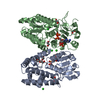
| ||||||||
|---|---|---|---|---|---|---|---|---|---|
| 1 |
| ||||||||
| Unit cell |
|
-Components
-Protein , 1 types, 2 molecules AB
| #1: Protein | Mass: 32146.008 Da / Num. of mol.: 2 Source method: isolated from a genetically manipulated source Source: (gene. exp.) ACINETOBACTER BAUMANNII ATCC 19606 (bacteria)Production host: References: UniProt: D0CBC8, methylenetetrahydrofolate dehydrogenase (NADP+), methenyltetrahydrofolate cyclohydrolase |
|---|
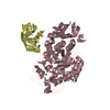
-Non-polymers , 5 types, 753 molecules 


| #2: Chemical |  Mass: 743.405 Da / Num. of mol.: 2 / Source method: obtained synthetically / Formula: C21H28N7O17P3 Mass: 743.405 Da / Num. of mol.: 2 / Source method: obtained synthetically / Formula: C21H28N7O17P3#3: Chemical | ChemComp-CL /  Mass: 35.453 Da / Num. of mol.: 5 / Source method: obtained synthetically / Formula: Cl Mass: 35.453 Da / Num. of mol.: 5 / Source method: obtained synthetically / Formula: Cl#4: Chemical | ChemComp-EDO /  Mass: 62.068 Da / Num. of mol.: 5 / Source method: obtained synthetically / Formula: C2H6O2 Mass: 62.068 Da / Num. of mol.: 5 / Source method: obtained synthetically / Formula: C2H6O2#5: Chemical | ChemComp-PEG / |  Mass: 106.120 Da / Num. of mol.: 1 / Source method: obtained synthetically / Formula: C4H10O3 Mass: 106.120 Da / Num. of mol.: 1 / Source method: obtained synthetically / Formula: C4H10O3#6: Water | ChemComp-HOH / | Mass: 18.015 Da / Num. of mol.: 740 / Source method: isolated from a natural source / Formula: H2O |
|---|
-Experimental details
-Experiment
| Experiment | Method: X-RAY DIFFRACTION / Number of used crystals: 1 |
|---|
- Sample preparation
| Crystal | Density Matthews: 2.4 Å3/Da / Density % sol: 49.2 % / Description: NONE |
|---|---|
| Crystal grow | Details: 0.1 M BIS-TRIS PH 5.5, 25 % PEG 3350, 0.2M MGCL AND 2 % DIOXANE |
-Data collection
| Diffraction | Mean temperature: 100 K |
|---|---|
| Diffraction source | Source: SYNCHROTRON / Site: ESRF  / Beamline: ID29 / Wavelength: 0.981 / Beamline: ID29 / Wavelength: 0.981 |
| Detector | Type: ADSC QUANTUM 315r / Detector: CCD / Date: May 25, 2009 |
| Radiation | Protocol: SINGLE WAVELENGTH / Monochromatic (M) / Laue (L): M / Scattering type: x-ray |
| Radiation wavelength | Wavelength: 0.981 Å / Relative weight: 1 |
| Reflection | Resolution: 1.45→52.6 Å / Num. obs: 100394 / % possible obs: 99.7 % / Observed criterion σ(I): 2 / Redundancy: 3.6 % / Biso Wilson estimate: 14.9 Å2 / Rmerge(I) obs: 0.06 / Net I/σ(I): 13.1 |
| Reflection shell | Resolution: 1.45→1.53 Å / Redundancy: 3.3 % / Rmerge(I) obs: 0.42 / Mean I/σ(I) obs: 2.9 / % possible all: 99.3 |
- Processing
| Software |
| ||||||||||||||||||||||||||||||||||||||||||||||||||||||||||||||||||||||||||||||||||||||||||||||||||||||||||||||||||||||||||||||||||||||||||||||||||||||||||||||||||||||||||||||||||||||
|---|---|---|---|---|---|---|---|---|---|---|---|---|---|---|---|---|---|---|---|---|---|---|---|---|---|---|---|---|---|---|---|---|---|---|---|---|---|---|---|---|---|---|---|---|---|---|---|---|---|---|---|---|---|---|---|---|---|---|---|---|---|---|---|---|---|---|---|---|---|---|---|---|---|---|---|---|---|---|---|---|---|---|---|---|---|---|---|---|---|---|---|---|---|---|---|---|---|---|---|---|---|---|---|---|---|---|---|---|---|---|---|---|---|---|---|---|---|---|---|---|---|---|---|---|---|---|---|---|---|---|---|---|---|---|---|---|---|---|---|---|---|---|---|---|---|---|---|---|---|---|---|---|---|---|---|---|---|---|---|---|---|---|---|---|---|---|---|---|---|---|---|---|---|---|---|---|---|---|---|---|---|---|---|
| Refinement | Method to determine structure: MOLECULAR REPLACEMENT Starting model: PDB ENTRY 1B0A Resolution: 1.45→52.6 Å / Cor.coef. Fo:Fc: 0.979 / Cor.coef. Fo:Fc free: 0.972 / SU B: 1.738 / SU ML: 0.03 / Cross valid method: THROUGHOUT / ESU R: 0.058 / ESU R Free: 0.052 / Stereochemistry target values: MAXIMUM LIKELIHOOD Details: HYDROGENS HAVE BEEN ADDED IN THE RIDING POSITIONS. U VALUES REFINED INDIVIDUALLY. RESIDUES WITH DISORDERED SIDE CHAINS ARE MODELED WITH AN OCCUPANCY OF 0.00. DISORDERED NICOTINAMIDE AND RIBOSE.
| ||||||||||||||||||||||||||||||||||||||||||||||||||||||||||||||||||||||||||||||||||||||||||||||||||||||||||||||||||||||||||||||||||||||||||||||||||||||||||||||||||||||||||||||||||||||
| Solvent computation | Ion probe radii: 0.8 Å / Shrinkage radii: 0.8 Å / VDW probe radii: 1.2 Å / Solvent model: MASK | ||||||||||||||||||||||||||||||||||||||||||||||||||||||||||||||||||||||||||||||||||||||||||||||||||||||||||||||||||||||||||||||||||||||||||||||||||||||||||||||||||||||||||||||||||||||
| Displacement parameters | Biso mean: 14.912 Å2
| ||||||||||||||||||||||||||||||||||||||||||||||||||||||||||||||||||||||||||||||||||||||||||||||||||||||||||||||||||||||||||||||||||||||||||||||||||||||||||||||||||||||||||||||||||||||
| Refinement step | Cycle: LAST / Resolution: 1.45→52.6 Å
| ||||||||||||||||||||||||||||||||||||||||||||||||||||||||||||||||||||||||||||||||||||||||||||||||||||||||||||||||||||||||||||||||||||||||||||||||||||||||||||||||||||||||||||||||||||||
| Refine LS restraints |
|