 Movie
Movie Controller


+ Open data

- Basic information
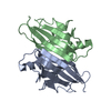
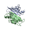
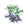
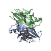
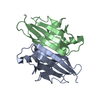
| Entry | Database: PDB / ID: 3kgs | ||||||
|---|---|---|---|---|---|---|---|
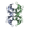
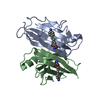
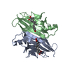
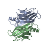
| Title | V30M mutant human transthyretin (TTR) (apoV30M) pH 7.5 | ||||||
 Components Components | Transthyretin | ||||||
 Keywords Keywords | TRANSPORT PROTEIN / TTR / TRANSTHYRETIN / AMYLOID / Amyloidosis / Hormone / Neuropathy / Polymorphism / Secreted / Thyroid hormone / Transport | ||||||
| Function / homology |  Function and homology information Function and homology informationRetinoid cycle disease events / thyroid hormone binding / The canonical retinoid cycle in rods (twilight vision) / Non-integrin membrane-ECM interactions / purine nucleobase metabolic process / Retinoid metabolism and transport / hormone activity / azurophil granule lumen / Amyloid fiber formation / Neutrophil degranulation ...Retinoid cycle disease events / thyroid hormone binding / The canonical retinoid cycle in rods (twilight vision) / Non-integrin membrane-ECM interactions / purine nucleobase metabolic process / Retinoid metabolism and transport / hormone activity / azurophil granule lumen / Amyloid fiber formation / Neutrophil degranulation / extracellular space / extracellular exosome / extracellular region / identical protein bindingSimilarity search - Function | ||||||
| Biological species |  Homo sapiens (human) Homo sapiens (human) | ||||||
| Method | X-RAY DIFFRACTION / SYNCHROTRON / MOLECULAR REPLACEMENT / Resolution: 1.8 Å | ||||||
 Authors Authors | Trivella, D.B. / Polikarpov, I. | ||||||
 Citation Citation | Journal: J.Struct.Biol. / Year: 2010 Title: Conformational differences between the wild type and V30M mutant transthyretin modulate its binding to genistein: implications to tetramer stability and ligand-binding. Authors: Trivella, D.B. / Bleicher, L. / Palmieri, L.C. / Wiggers, H.J. / Montanari, C.A. / Kelly, J.W. / Lima, L.M. / Foguel, D. / Polikarpov, I. | ||||||
| History |
|
- Structure visualization
| Structure viewer | Molecule: MolmilJmol/JSmol |
|---|
- Downloads & links
-Download
| PDBx/mmCIF format | 3kgs.cif.gz | 63.8 KB | Display | PDBx/mmCIF format |
|---|---|---|---|---|
| PDB format | pdb3kgs.ent.gz | 48.1 KB | Display | PDB format |
| PDBx/mmJSON format | 3kgs.json.gz | Tree view | PDBx/mmJSON format | |
| Others |  Other downloads Other downloads |
-Validation report
| Arichive directory | https://data.pdbj.org/pub/pdb/validation_reports/kg/3kgsftp://data.pdbj.org/pub/pdb/validation_reports/kg/3kgs | HTTPS FTP |
|---|
-Related structure data
| Related structure data | 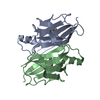 3kgtC 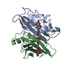 3kguC 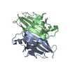 1f41S C: citing same article ( S: Starting model for refinement |
|---|---|
| Similar structure data |
-Links
 PDBj
PDBj


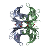
- Assembly
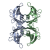
| Deposited unit | 
| |||||||||
|---|---|---|---|---|---|---|---|---|---|---|
| 1 | 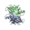
| |||||||||
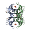
| Unit cell |
| |||||||||
| Components on special symmetry positions |
|
-Components
| #1: Protein | / Prealbumin / TBPA / TTR / ATTR Mass: 13809.426 Da / Num. of mol.: 2 / Fragment: UNP residues 20-147 / Mutation: V30M Source method: isolated from a genetically manipulated source Source: (gene. exp.) Homo sapiens (human) / Gene: PALB, TTR / Production host:  Escherichia coli (E. coli) / Strain (production host): BL21(DE3) / References: UniProt: P02766 Escherichia coli (E. coli) / Strain (production host): BL21(DE3) / References: UniProt: P02766#2: Water | ChemComp-HOH / | Water Mass: 18.015 Da / Num. of mol.: 207 / Source method: isolated from a natural source / Formula: H2O Mass: 18.015 Da / Num. of mol.: 207 / Source method: isolated from a natural source / Formula: H2O |
|---|
-Experimental details
-Experiment
| Experiment | Method: X-RAY DIFFRACTION / Number of used crystals: 1 |
|---|
- Sample preparation
| Crystal | Density Matthews: 2.29 Å3/Da / Density % sol: 46.32 % |
|---|---|
| Crystal grow | Temperature: 293 K / Method: vapor diffusion, hanging drop / pH: 7.5 Details: 0.2 M CaCl2, 0.1 M HEPES pH 7.5, 28% PEG 400, vapor diffusion, hanging drop, temperature 293K |
-Data collection
| Diffraction | Mean temperature: 100 K |
|---|---|
| Diffraction source | Source: SYNCHROTRON / Site: LNLS  / Beamline: W01B-MX2 / Wavelength: 1.4586 Å / Beamline: W01B-MX2 / Wavelength: 1.4586 Å |
| Detector | Type: MARMOSAIC 225 mm CCD / Detector: CCD / Date: Mar 15, 2009 |
| Radiation | Protocol: SINGLE WAVELENGTH / Monochromatic (M) / Laue (L): M / Scattering type: x-ray |
| Radiation wavelength | Wavelength: 1.4586 Å / Relative weight: 1 |
| Reflection | Resolution: 1.8→30 Å / Num. obs: 26041 / % possible obs: 97 % / Redundancy: 3.7 % / Rmerge(I) obs: 0.063 |
| Reflection shell | Resolution: 1.8→1.84 Å / Rmerge(I) obs: 0.063 / Mean I/σ(I) obs: 7.1 |
- Processing
| Software |
| ||||||||||||||||||||||||||||||||||||||||||||||||||||||||||||||||||||||||||||||||||||||||||
|---|---|---|---|---|---|---|---|---|---|---|---|---|---|---|---|---|---|---|---|---|---|---|---|---|---|---|---|---|---|---|---|---|---|---|---|---|---|---|---|---|---|---|---|---|---|---|---|---|---|---|---|---|---|---|---|---|---|---|---|---|---|---|---|---|---|---|---|---|---|---|---|---|---|---|---|---|---|---|---|---|---|---|---|---|---|---|---|---|---|---|---|
| Refinement | Method to determine structure: MOLECULAR REPLACEMENT Starting model: PDB entry 1F41 Resolution: 1.8→22.1 Å / Cor.coef. Fo:Fc: 0.948 / Cor.coef. Fo:Fc free: 0.927 / WRfactor Rfree: 0.25 / WRfactor Rwork: 0.203 / Occupancy max: 1 / Occupancy min: 0.25 / FOM work R set: 0.83 / SU B: 2.989 / SU ML: 0.095 / SU R Cruickshank DPI: 0.153 / SU Rfree: 0.146 / Cross valid method: THROUGHOUT / σ(F): 0 / ESU R: 0.153 / ESU R Free: 0.146 / Stereochemistry target values: MAXIMUM LIKELIHOOD / Details: HYDROGENS HAVE BEEN ADDED IN THE RIDING POSITIONS
| ||||||||||||||||||||||||||||||||||||||||||||||||||||||||||||||||||||||||||||||||||||||||||
| Solvent computation | Ion probe radii: 0.8 Å / Shrinkage radii: 0.8 Å / VDW probe radii: 1.2 Å / Solvent model: MASK | ||||||||||||||||||||||||||||||||||||||||||||||||||||||||||||||||||||||||||||||||||||||||||
| Displacement parameters | Biso max: 70.12 Å2 / Biso mean: 20.451 Å2 / Biso min: 7.47 Å2
| ||||||||||||||||||||||||||||||||||||||||||||||||||||||||||||||||||||||||||||||||||||||||||
| Refinement step | Cycle: LAST / Resolution: 1.8→22.1 Å
| ||||||||||||||||||||||||||||||||||||||||||||||||||||||||||||||||||||||||||||||||||||||||||
| Refine LS restraints |
| ||||||||||||||||||||||||||||||||||||||||||||||||||||||||||||||||||||||||||||||||||||||||||
| LS refinement shell | Resolution: 1.8→1.846 Å / Total num. of bins used: 20
|
