 Movie
Movie Controller

[English] 日本語
 Yorodumi
- PDB-3fgs: Crystal structure of G65R/K206E double mutant of the N-lobe human... -
+ Open data

- Basic information
| Entry | Database: PDB / ID: 3fgs | ||||||
|---|---|---|---|---|---|---|---|
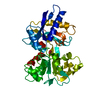
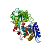
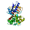
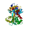
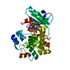
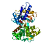
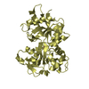
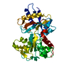
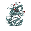
| Title | Crystal structure of G65R/K206E double mutant of the N-lobe human transferrin | ||||||
 Components Components | Serotransferrin | ||||||
 Keywords Keywords | METAL TRANSPORT /  Human transferrin / Iron binding protein / Dilysine pair / Disease mutation / Glycoprotein / Ion transport / Iron / Iron transport / Metal-binding / Methylation / Phosphoprotein / Polymorphism / Secreted / Transport Human transferrin / Iron binding protein / Dilysine pair / Disease mutation / Glycoprotein / Ion transport / Iron / Iron transport / Metal-binding / Methylation / Phosphoprotein / Polymorphism / Secreted / Transport | ||||||
| Function / homology |  Function and homology information Function and homology informationiron chaperone activity / Transferrin endocytosis and recycling / transferrin receptor binding / basal part of cell / positive regulation of cell motility / endocytic vesicle / positive regulation of bone resorption / positive regulation of phosphorylation / clathrin-coated pit / ERK1 and ERK2 cascade ...iron chaperone activity / Transferrin endocytosis and recycling / transferrin receptor binding / basal part of cell / positive regulation of cell motility / endocytic vesicle / positive regulation of bone resorption / positive regulation of phosphorylation / clathrin-coated pit / ERK1 and ERK2 cascade / osteoclast differentiation / basal plasma membrane / ferric iron binding / actin filament organization / ferrous iron binding / Post-translational protein phosphorylation / Iron uptake and transport / clathrin-coated endocytic vesicle membrane / regulation of protein stability / regulation of iron ion transport / HFE-transferrin receptor complex / cellular response to iron ion / recycling endosome / positive regulation of receptor-mediated endocytosis / Regulation of Insulin-like Growth Factor (IGF) transport and uptake by Insulin-like Growth Factor Binding Proteins (IGFBPs) / late endosome / Cargo recognition for clathrin-mediated endocytosis / Platelet degranulation / Clathrin-mediated endocytosis / antibacterial humoral response / iron ion transport / cytoplasmic vesicle / secretory granule lumen / blood microparticle / vesicle / intracellular iron ion homeostasis / early endosome / endosome membrane / apical plasma membrane / endoplasmic reticulum lumen / perinuclear region of cytoplasm / positive regulation of DNA-templated transcription / cell surface / extracellular space / extracellular exosome / extracellular region / plasma membraneSimilarity search - Function | ||||||
| Biological species |  Homo sapiens (human) Homo sapiens (human) | ||||||
| Method | X-RAY DIFFRACTION / MOLECULAR REPLACEMENT / molecular replacement / Resolution: 1.8 Å | ||||||
 Authors Authors | Halbrooks, P.J. / Mason, A.B. / Everse, S.J. | ||||||
 Citation Citation | Journal: Biochemistry / Year: 2009 Title: Structural and Functional Consequences of the Substitution of Glycine 65 with Arginine in the N-Lobe of Human Transferrin. Authors: Mason, A.B. / Halbrooks, P.J. / James, N.G. / Byrne, S.L. / Grady, J.K. / Chasteen, N.D. / Bobst, C.E. / Kaltashov, I.A. / Smith, V.C. / Macgillivray, R.T. / Everse, S.J. | ||||||
| History |
|
- Structure visualization
| Structure viewer | Molecule: MolmilJmol/JSmol |
|---|
- Downloads & links
-Download
| PDBx/mmCIF format | 3fgs.cif.gz | 80.1 KB | Display | PDBx/mmCIF format |
|---|---|---|---|---|
| PDB format | pdb3fgs.ent.gz | 63.1 KB | Display | PDB format |
| PDBx/mmJSON format | 3fgs.json.gz | Tree view | PDBx/mmJSON format | |
| Others |  Other downloads Other downloads |
-Validation report
| Arichive directory | https://data.pdbj.org/pub/pdb/validation_reports/fg/3fgsftp://data.pdbj.org/pub/pdb/validation_reports/fg/3fgs | HTTPS FTP |
|---|
-Related structure data
| Related structure data | |
|---|---|
| Similar structure data |


-Links
 PDBj
PDBj


- Assembly
| Deposited unit | 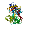
| ||||||||
|---|---|---|---|---|---|---|---|---|---|
| 1 |
| ||||||||
| Unit cell |
|
-Components
| #1: Protein | Mass: 37315.344 Da / Num. of mol.: 1 / Fragment: Peptidase S60 1 domain / Mutation: G65R, K206E Source method: isolated from a genetically manipulated source Source: (gene. exp.) Homo sapiens (human) / Gene: PRO1400, TF, transferrin / Plasmid: pNUT / Production host:  Mesocricetus auratus (golden hamster) / Strain (production host): BHK / References: UniProt: P02787 Mesocricetus auratus (golden hamster) / Strain (production host): BHK / References: UniProt: P02787 |
|---|---|
| #2: Chemical | ChemComp-CO3 / Carbonate  Mass: 60.009 Da / Num. of mol.: 1 / Source method: obtained synthetically / Formula: CO3 Mass: 60.009 Da / Num. of mol.: 1 / Source method: obtained synthetically / Formula: CO3 |
| #3: Chemical | ChemComp-FE / Iron  Mass: 55.845 Da / Num. of mol.: 1 / Source method: obtained synthetically / Formula: Fe Mass: 55.845 Da / Num. of mol.: 1 / Source method: obtained synthetically / Formula: Fe |
| #4: Water | ChemComp-HOH / Water Mass: 18.015 Da / Num. of mol.: 269 / Source method: isolated from a natural source / Formula: H2O Mass: 18.015 Da / Num. of mol.: 269 / Source method: isolated from a natural source / Formula: H2O |
-Experimental details
-Experiment
| Experiment | Method: X-RAY DIFFRACTION / Number of used crystals: 1 |
|---|
- Sample preparation
| Crystal | Density Matthews: 2.22 Å3/Da / Density % sol: 44.49 % |
|---|---|
| Crystal grow | Temperature: 293 K / Method: vapor diffusion, hanging drop / pH: 7.7 Details: 200 mM potassium acetate buffer (pH 7.7) containing 10 mM KCl and 18% polyethylene glycol 3350. Concentration of the mutant was 17.5 mg/mL. Crystals appeared in approximately a week ...Details: 200 mM potassium acetate buffer (pH 7.7) containing 10 mM KCl and 18% polyethylene glycol 3350. Concentration of the mutant was 17.5 mg/mL. Crystals appeared in approximately a week following micro-seeding with wild-type N-lobe, VAPOR DIFFUSION, HANGING DROP, temperature 293K |
-Data collection
| Diffraction | Mean temperature: 100 K | |||||||||||||||||||||||||||||||||||||||||||||||||||||||
|---|---|---|---|---|---|---|---|---|---|---|---|---|---|---|---|---|---|---|---|---|---|---|---|---|---|---|---|---|---|---|---|---|---|---|---|---|---|---|---|---|---|---|---|---|---|---|---|---|---|---|---|---|---|---|---|---|
| Diffraction source | Source: ROTATING ANODE / Type: RIGAKU RU300 / Wavelength: 1.5418 Å | |||||||||||||||||||||||||||||||||||||||||||||||||||||||
| Detector | Type: MAR scanner 345 mm plate / Detector: IMAGE PLATE / Date: Aug 7, 2001 / Details: Xenocs FOX-2D Multilayer Mirrors | |||||||||||||||||||||||||||||||||||||||||||||||||||||||
| Radiation | Protocol: SINGLE WAVELENGTH / Monochromatic (M) / Laue (L): M / Scattering type: x-ray | |||||||||||||||||||||||||||||||||||||||||||||||||||||||
| Radiation wavelength | Wavelength: 1.5418 Å / Relative weight: 1 | |||||||||||||||||||||||||||||||||||||||||||||||||||||||
| Reflection | Resolution: 1.8→30 Å / Num. obs: 29637 / % possible obs: 93.3 % / Rmerge(I) obs: 0.049 / Χ2: 1.511 | |||||||||||||||||||||||||||||||||||||||||||||||||||||||
| Reflection shell |
|
-Phasing
| Phasing | Method: molecular replacement |
|---|---|
| Phasing MR | Method rotation: fast direct / Method translation: &STRIP%trans_method |
- Processing
| Software |
| ||||||||||||||||||||||||||||
|---|---|---|---|---|---|---|---|---|---|---|---|---|---|---|---|---|---|---|---|---|---|---|---|---|---|---|---|---|---|
| Refinement | Method to determine structure: MOLECULAR REPLACEMENT / Resolution: 1.8→19.49 Å / Occupancy max: 1 / Occupancy min: 1 / σ(F): 0
| ||||||||||||||||||||||||||||
| Solvent computation | Bsol: 36.137 Å2 | ||||||||||||||||||||||||||||
| Displacement parameters | Biso max: 45.94 Å2 / Biso mean: 14.999 Å2 / Biso min: 5.79 Å2
| ||||||||||||||||||||||||||||
| Refinement step | Cycle: LAST / Resolution: 1.8→19.49 Å
| ||||||||||||||||||||||||||||
| Refine LS restraints |
| ||||||||||||||||||||||||||||
| Xplor file |
|
