 Movie
Movie Controller

[English] 日本語
 Yorodumi
- PDB-1fqe: CRYSTAL STRUCTURES OF MUTANT (K206A) THAT ABOLISH THE DILYSINE IN... -
+ Open data

- Basic information
| Entry | Database: PDB / ID: 1fqe | ||||||
|---|---|---|---|---|---|---|---|
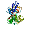
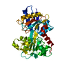
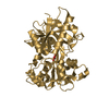
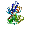
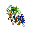
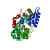
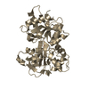
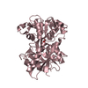
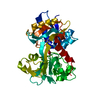
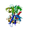
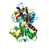
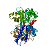
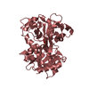
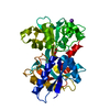
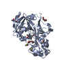
| Title | CRYSTAL STRUCTURES OF MUTANT (K206A) THAT ABOLISH THE DILYSINE INTERACTION IN THE N-LOBE OF HUMAN TRANSFERRIN | ||||||
 Components Components | SEROTRANSFERRIN | ||||||
 Keywords Keywords | METAL TRANSPORT / IRON TRANSPORT / TRANSFERRIN / N-LOBE / IRON-RELEASE / DILYSINE INTERACTION | ||||||
| Function / homology |  Function and homology information Function and homology informationiron chaperone activity / transferrin receptor binding / Transferrin endocytosis and recycling / basal part of cell / endocytic vesicle / clathrin-coated pit / ferric iron binding / osteoclast differentiation / basal plasma membrane / Post-translational protein phosphorylation ...iron chaperone activity / transferrin receptor binding / Transferrin endocytosis and recycling / basal part of cell / endocytic vesicle / clathrin-coated pit / ferric iron binding / osteoclast differentiation / basal plasma membrane / Post-translational protein phosphorylation / iron ion transport / clathrin-coated endocytic vesicle membrane / HFE-transferrin receptor complex / regulation of protein stability / cellular response to iron ion / ferrous iron binding / Iron uptake and transport / positive regulation of receptor-mediated endocytosis / multicellular organismal-level iron ion homeostasis / recycling endosome / Regulation of Insulin-like Growth Factor (IGF) transport and uptake by Insulin-like Growth Factor Binding Proteins (IGFBPs) / late endosome / Platelet degranulation / Cargo recognition for clathrin-mediated endocytosis / positive regulation of proteasomal ubiquitin-dependent protein catabolic process / antibacterial humoral response / Clathrin-mediated endocytosis / cytoplasmic vesicle / secretory granule lumen / blood microparticle / vesicle / intracellular iron ion homeostasis / transmembrane transporter binding / early endosome / cell surface receptor signaling pathway / endosome membrane / apical plasma membrane / endoplasmic reticulum lumen / perinuclear region of cytoplasm / enzyme binding / cell surface / : / extracellular exosome / extracellular region / plasma membrane Similarity search - Function | ||||||
| Biological species |  Homo sapiens (human) Homo sapiens (human) | ||||||
| Method |  X-RAY DIFFRACTION / SYNCHROTRON / Resolution: 1.8 Å X-RAY DIFFRACTION / SYNCHROTRON / Resolution: 1.8 Å | ||||||
 Authors Authors | Nurizzo, D. / Baker, H.M. / Baker, E.N. | ||||||
 Citation Citation | Journal: Biochemistry / Year: 2001 Title: Crystal structures and iron release properties of mutants (K206A and K296A) that abolish the dilysine interaction in the N-lobe of human transferrin. Authors: Nurizzo, D. / Baker, H.M. / He, Q.Y. / MacGillivray, R.T. / Mason, A.B. / Woodworth, R.C. / Baker, E.N. | ||||||
| History |
|
- Structure visualization
| Structure viewer | Molecule: MolmilJmol/JSmol |
|---|
- Downloads & links
-Download
| PDBx/mmCIF format | 1fqe.cif.gz | 88.2 KB | Display | PDBx/mmCIF format |
|---|---|---|---|---|
| PDB format | pdb1fqe.ent.gz | 65.5 KB | Display | PDB format |
| PDBx/mmJSON format | 1fqe.json.gz | Tree view | PDBx/mmJSON format | |
| Others |  Other downloads Other downloads |
-Validation report
| Arichive directory | https://data.pdbj.org/pub/pdb/validation_reports/fq/1fqeftp://data.pdbj.org/pub/pdb/validation_reports/fq/1fqe | HTTPS FTP |
|---|
-Related structure data


-Links
 PDBj
PDBj


- Assembly
| Deposited unit | 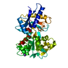
| ||||||||
|---|---|---|---|---|---|---|---|---|---|
| 1 |
| ||||||||
| Unit cell |
| ||||||||
| Details | The biological assembly is a monomer constituted by two domains in between the iron and its carbonate counterpart are linked. |
-Components
| #1: Protein | Mass: 36546.559 Da / Num. of mol.: 1 / Fragment: N-LOBE / Mutation: K206A Source method: isolated from a genetically manipulated source Source: (gene. exp.) Homo sapiens (human) / Tissue fraction: SERUM / Plasmid: PNUT-BHK / Production host:  Mesocricetus auratus (golden hamster) / References: UniProt: P02787 Mesocricetus auratus (golden hamster) / References: UniProt: P02787 |
|---|---|
| #2: Chemical | ChemComp-FE /   Mass: 55.845 Da / Num. of mol.: 1 / Source method: obtained synthetically / Formula: Fe Mass: 55.845 Da / Num. of mol.: 1 / Source method: obtained synthetically / Formula: Fe |
| #3: Chemical | ChemComp-CO3 /   Mass: 60.009 Da / Num. of mol.: 1 / Source method: obtained synthetically / Formula: CO3 Mass: 60.009 Da / Num. of mol.: 1 / Source method: obtained synthetically / Formula: CO3 |
| #4: Chemical | ChemComp-K /   Mass: 39.098 Da / Num. of mol.: 1 / Source method: obtained synthetically / Formula: K Mass: 39.098 Da / Num. of mol.: 1 / Source method: obtained synthetically / Formula: K |
| #5: Water | ChemComp-HOH /  Mass: 18.015 Da / Num. of mol.: 442 / Source method: isolated from a natural source / Formula: H2O Mass: 18.015 Da / Num. of mol.: 442 / Source method: isolated from a natural source / Formula: H2O |
| Has protein modification | Y |
-Experimental details
-Experiment
| Experiment | Method: X-RAY DIFFRACTION / Number of used crystals: 1 |
|---|
- Sample preparation
| Crystal | Density Matthews: 2.34 Å3/Da / Density % sol: 47.38 % | |||||||||||||||||||||||||
|---|---|---|---|---|---|---|---|---|---|---|---|---|---|---|---|---|---|---|---|---|---|---|---|---|---|---|
| Crystal grow | Temperature: 293 K / Method: vapor diffusion, hanging drop / pH: 7.4 Details: 20-25% PolyEthyleneGlycol 3350, 100mM Potassium acetate, pH 7.4, VAPOR DIFFUSION, HANGING DROP, temperature 293K | |||||||||||||||||||||||||
| Crystal grow | *PLUS | |||||||||||||||||||||||||
| Components of the solutions | *PLUS
|
-Data collection
| Diffraction | Mean temperature: 110 K |
|---|---|
| Diffraction source | Source: SYNCHROTRON / Site: SSRL  / Beamline: BL7-1 / Wavelength: 1.08 / Beamline: BL7-1 / Wavelength: 1.08 |
| Detector | Type: MARRESEARCH / Detector: IMAGE PLATE / Date: Feb 11, 2000 |
| Radiation | Protocol: SINGLE WAVELENGTH / Monochromatic (M) / Laue (L): M / Scattering type: x-ray |
| Radiation wavelength | Wavelength: 1.08 Å / Relative weight: 1 |
| Reflection | Resolution: 1.8→14.73 Å / Num. all: 373302 / Num. obs: 29721 / % possible obs: 91.2 % / Observed criterion σ(F): 0 / Observed criterion σ(I): 2.4 / Redundancy: 3.1 % / Biso Wilson estimate: 9 Å2 / Rmerge(I) obs: 0.071 / Net I/σ(I): 7.4 |
| Reflection shell | Resolution: 1.8→1.9 Å / Redundancy: 2.8 % / Rmerge(I) obs: 0.185 / Num. unique all: 10127 / % possible all: 82.1 |
| Reflection | *PLUS Num. measured all: 373302 |
| Reflection shell | *PLUS % possible obs: 82.1 % |
- Processing
| Software |
| ||||||||||||||||||||||||||||||||||||||||
|---|---|---|---|---|---|---|---|---|---|---|---|---|---|---|---|---|---|---|---|---|---|---|---|---|---|---|---|---|---|---|---|---|---|---|---|---|---|---|---|---|---|
| Refinement | Resolution: 1.8→14.73 Å / Rfactor Rfree error: 0.007 / Cross valid method: THROUGHOUT / σ(F): 0 / σ(I): 0 / Stereochemistry target values: Engh & Huber
| ||||||||||||||||||||||||||||||||||||||||
| Solvent computation | Solvent model: FLAT MODEL / Bsol: 24.0949 Å2 / ksol: 0.288978 e/Å3 | ||||||||||||||||||||||||||||||||||||||||
| Displacement parameters | Biso mean: 17.7 Å2
| ||||||||||||||||||||||||||||||||||||||||
| Refine analyze |
| ||||||||||||||||||||||||||||||||||||||||
| Refinement step | Cycle: LAST / Resolution: 1.8→14.73 Å
| ||||||||||||||||||||||||||||||||||||||||
| Refine LS restraints |
| ||||||||||||||||||||||||||||||||||||||||
| LS refinement shell | Resolution: 1.8→1.91 Å / Rfactor Rfree error: 0.019 / Total num. of bins used: 6
| ||||||||||||||||||||||||||||||||||||||||
| Xplor file |
| ||||||||||||||||||||||||||||||||||||||||
| Software | *PLUS Name: CNS / Version: 1 / Classification: refinement | ||||||||||||||||||||||||||||||||||||||||
| Refinement | *PLUS Highest resolution: 1.8 Å / σ(F): 0 / % reflection Rfree: 4 % | ||||||||||||||||||||||||||||||||||||||||
| Solvent computation | *PLUS | ||||||||||||||||||||||||||||||||||||||||
| Displacement parameters | *PLUS Biso mean: 17.7 Å2 | ||||||||||||||||||||||||||||||||||||||||
| Refine LS restraints | *PLUS
| ||||||||||||||||||||||||||||||||||||||||
| LS refinement shell | *PLUS Rfactor Rfree: 0.251 / % reflection Rfree: 3.8 % / Rfactor Rwork: 0.252 |
