 Movie
Movie Controller

[English] 日本語
 Yorodumi
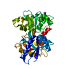
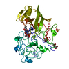
- PDB-1oqh: Crystal Structure of the R124A mutant of the N-lobe human transferrin -
+ Open data

- Basic information
| Entry | Database: PDB / ID: 1oqh | ||||||
|---|---|---|---|---|---|---|---|
| Title | Crystal Structure of the R124A mutant of the N-lobe human transferrin | ||||||
 Components Components | Serotransferrin | ||||||
 Keywords Keywords | TRANSPORT PROTEIN / transferrin mutagenesis / iron binding / anion binding | ||||||
| Function / homology |  Function and homology information Function and homology informationiron chaperone activity / transferrin receptor binding / Transferrin endocytosis and recycling / basal part of cell / endocytic vesicle / clathrin-coated pit / ferric iron binding / osteoclast differentiation / basal plasma membrane / Post-translational protein phosphorylation ...iron chaperone activity / transferrin receptor binding / Transferrin endocytosis and recycling / basal part of cell / endocytic vesicle / clathrin-coated pit / ferric iron binding / osteoclast differentiation / basal plasma membrane / Post-translational protein phosphorylation / iron ion transport / clathrin-coated endocytic vesicle membrane / HFE-transferrin receptor complex / regulation of protein stability / cellular response to iron ion / ferrous iron binding / Iron uptake and transport / positive regulation of receptor-mediated endocytosis / multicellular organismal-level iron ion homeostasis / recycling endosome / Regulation of Insulin-like Growth Factor (IGF) transport and uptake by Insulin-like Growth Factor Binding Proteins (IGFBPs) / late endosome / Platelet degranulation / Cargo recognition for clathrin-mediated endocytosis / positive regulation of proteasomal ubiquitin-dependent protein catabolic process / antibacterial humoral response / Clathrin-mediated endocytosis / cytoplasmic vesicle / secretory granule lumen / blood microparticle / vesicle / intracellular iron ion homeostasis / transmembrane transporter binding / early endosome / cell surface receptor signaling pathway / endosome membrane / apical plasma membrane / endoplasmic reticulum lumen / perinuclear region of cytoplasm / enzyme binding / cell surface / : / extracellular exosome / extracellular region / plasma membrane Similarity search - Function | ||||||
| Biological species |  Homo sapiens (human) Homo sapiens (human) | ||||||
| Method |  X-RAY DIFFRACTION / MOLECULAR REPLACEMENT / Resolution: 2.4 Å X-RAY DIFFRACTION / MOLECULAR REPLACEMENT / Resolution: 2.4 Å | ||||||
 Authors Authors | Baker, H.M. / He, Q.-Y. / Brigg, S.K. / Mason, A.B. / N Baker, E. | ||||||
 Citation Citation | Journal: Biochemistry / Year: 2003 Title: Structural and functional consequences of binding site mutations in transferrin: crystal structures of the Asp63Glu and Arg124Ala mutants of the N-lobe of human transferrin Authors: Baker, H.M. / He, Q.-Y. / Briggs, S.K. / Mason, A.B. / Baker, E.N. | ||||||
| History |
|
- Structure visualization
| Structure viewer | Molecule: MolmilJmol/JSmol |
|---|
- Downloads & links
-Download
| PDBx/mmCIF format | 1oqh.cif.gz | 78.9 KB | Display | PDBx/mmCIF format |
|---|---|---|---|---|
| PDB format | pdb1oqh.ent.gz | 58.5 KB | Display | PDB format |
| PDBx/mmJSON format | 1oqh.json.gz | Tree view | PDBx/mmJSON format | |
| Others |  Other downloads Other downloads |
-Validation report
| Arichive directory | https://data.pdbj.org/pub/pdb/validation_reports/oq/1oqhftp://data.pdbj.org/pub/pdb/validation_reports/oq/1oqh | HTTPS FTP |
|---|
-Related structure data
| Related structure data |  1oqgC  1a8eS S: Starting model for refinement C: citing same article ( |
|---|---|
| Similar structure data |


-Links
 PDBj
PDBj


- Assembly
| Deposited unit | 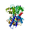
| ||||||||
|---|---|---|---|---|---|---|---|---|---|
| 1 |
| ||||||||
| Unit cell |
|
-Components
| #1: Protein | Mass: 37129.152 Da / Num. of mol.: 1 / Fragment: N-lobe / Mutation: R124A Source method: isolated from a genetically manipulated source Source: (gene. exp.) Homo sapiens (human) / Plasmid: pNUT / Cell (production host): baby hamster kidney (BHK) CELLS / Production host:  Cricetinae gen. sp. (mammal) / References: UniProt: P02787 Cricetinae gen. sp. (mammal) / References: UniProt: P02787 |
|---|---|
| #2: Chemical | ChemComp-CO3 /   Mass: 60.009 Da / Num. of mol.: 1 / Source method: obtained synthetically / Formula: CO3 Mass: 60.009 Da / Num. of mol.: 1 / Source method: obtained synthetically / Formula: CO3 |
| #3: Chemical | ChemComp-FE /   Mass: 55.845 Da / Num. of mol.: 1 / Source method: obtained synthetically / Formula: Fe Mass: 55.845 Da / Num. of mol.: 1 / Source method: obtained synthetically / Formula: Fe |
| #4: Chemical | ChemComp-K /   Mass: 39.098 Da / Num. of mol.: 1 / Source method: obtained synthetically / Formula: K Mass: 39.098 Da / Num. of mol.: 1 / Source method: obtained synthetically / Formula: K |
| #5: Water | ChemComp-HOH /  Mass: 18.015 Da / Num. of mol.: 140 / Source method: isolated from a natural source / Formula: H2O Mass: 18.015 Da / Num. of mol.: 140 / Source method: isolated from a natural source / Formula: H2O |
| Has protein modification | Y |
-Experimental details
-Experiment
| Experiment | Method: X-RAY DIFFRACTION / Number of used crystals: 1 |
|---|
- Sample preparation
| Crystal | Density Matthews: 2.25 Å3/Da / Density % sol: 45 % | ||||||||||||||||||||||||||||||
|---|---|---|---|---|---|---|---|---|---|---|---|---|---|---|---|---|---|---|---|---|---|---|---|---|---|---|---|---|---|---|---|
| Crystal grow | Temperature: 291 K / Method: vapor diffusion, hanging drop / pH: 7.4 Details: PEG 3350, potassium acetate, ammonium bicarbonate, pH 7.4, VAPOR DIFFUSION, HANGING DROP, temperature 291K | ||||||||||||||||||||||||||||||
| Crystal grow | *PLUS Temperature: 18 ℃ / Details: used microseeding | ||||||||||||||||||||||||||||||
| Components of the solutions | *PLUS
|
-Data collection
| Diffraction | Mean temperature: 113 K |
|---|---|
| Diffraction source | Source: ROTATING ANODE / Type: RIGAKU RU300 / Wavelength: 1.5418 Å |
| Detector | Type: MARRESEARCH / Detector: AREA DETECTOR / Date: May 12, 2002 / Details: osmic confocal |
| Radiation | Protocol: SINGLE WAVELENGTH / Monochromatic (M) / Laue (L): M / Scattering type: x-ray |
| Radiation wavelength | Wavelength: 1.5418 Å / Relative weight: 1 |
| Reflection | Resolution: 2.4→30 Å / Num. all: 12875 / Num. obs: 12186 / % possible obs: 91.5 % / Observed criterion σ(F): 0 / Observed criterion σ(I): 0 / Redundancy: 3.3 % / Rmerge(I) obs: 0.1 / Net I/σ(I): 7.4 |
| Reflection shell | Resolution: 2.4→2.51 Å / Rmerge(I) obs: 0.43 / Mean I/σ(I) obs: 2.2 / Num. unique all: 1377 / % possible all: 91.4 |
| Reflection | *PLUS Num. obs: 12875 / Rmerge(I) obs: 0.1 |
| Reflection shell | *PLUS Highest resolution: 2.4 Å / % possible obs: 91.4 % / Redundancy: 2.9 % / Rmerge(I) obs: 0.43 |
- Processing
| Software |
| ||||||||||||||||||||||||||||||||||||||||||||||||||||||||||||
|---|---|---|---|---|---|---|---|---|---|---|---|---|---|---|---|---|---|---|---|---|---|---|---|---|---|---|---|---|---|---|---|---|---|---|---|---|---|---|---|---|---|---|---|---|---|---|---|---|---|---|---|---|---|---|---|---|---|---|---|---|---|
| Refinement | Method to determine structure: MOLECULAR REPLACEMENT Starting model: PDB ENTRY 1A8E Resolution: 2.4→30 Å / Isotropic thermal model: anisotropic / Cross valid method: THROUGHOUT / σ(F): 0 / σ(I): 0 / Stereochemistry target values: Engh & Huber
| ||||||||||||||||||||||||||||||||||||||||||||||||||||||||||||
| Displacement parameters |
| ||||||||||||||||||||||||||||||||||||||||||||||||||||||||||||
| Refinement step | Cycle: LAST / Resolution: 2.4→30 Å
| ||||||||||||||||||||||||||||||||||||||||||||||||||||||||||||
| Refine LS restraints |
| ||||||||||||||||||||||||||||||||||||||||||||||||||||||||||||
| LS refinement shell | Resolution: 2.4→2.47 Å
| ||||||||||||||||||||||||||||||||||||||||||||||||||||||||||||
| Xplor file |
|
