 Movie
Movie Controller

[English] 日本語
 Yorodumi
- PDB-3bez: Crystal structure of Escherichia coli Signal peptide peptidase (S... -
+ Open data

- Basic information
| Entry | Database: PDB / ID: 3bez | ||||||
|---|---|---|---|---|---|---|---|
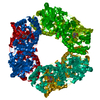
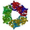
| Title | Crystal structure of Escherichia coli Signal peptide peptidase (SppA), SeMet crystals | ||||||
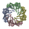
 Components Components | Protease 4 | ||||||
 Keywords Keywords | HYDROLASE / protease / bacterial / Inner membrane / Membrane / Transmembrane | ||||||
| Function / homology |  Function and homology information Function and homology informationsignal peptide processing / Hydrolases; Acting on peptide bonds (peptidases); Serine endopeptidases / serine-type peptidase activity / endopeptidase activity / membrane / plasma membraneSimilarity search - Function | ||||||
| Biological species |  Escherichia coli (E. coli) Escherichia coli (E. coli) | ||||||
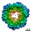
| Method | X-RAY DIFFRACTION / SYNCHROTRON / SAD / Resolution: 2.76 Å | ||||||
 Authors Authors | Paetzel, M. | ||||||
 Citation Citation | Journal: J.Mol.Biol. / Year: 2008 Title: Crystal structure of a bacterial signal Peptide peptidase. Authors: Kim, A.C. / Oliver, D.C. / Paetzel, M. | ||||||
| History |
|
- Structure visualization
| Structure viewer | Molecule: MolmilJmol/JSmol |
|---|
- Downloads & links
-Download
| PDBx/mmCIF format | 3bez.cif.gz | 374.1 KB | Display | PDBx/mmCIF format |
|---|---|---|---|---|
| PDB format | pdb3bez.ent.gz | 313.1 KB | Display | PDB format |
| PDBx/mmJSON format | 3bez.json.gz | Tree view | PDBx/mmJSON format | |
| Others |  Other downloads Other downloads |
-Validation report
| Arichive directory | https://data.pdbj.org/pub/pdb/validation_reports/be/3bezftp://data.pdbj.org/pub/pdb/validation_reports/be/3bez | HTTPS FTP |
|---|
-Related structure data


-Links
 PDBj
PDBj- Assembly
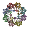
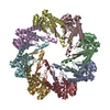
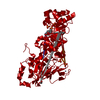
| Deposited unit | 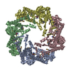
| |||||||||||||||||||||||||||||||||||||||||||||||||||||||||||||||||||||||||||||||||||||||||||||||||||
|---|---|---|---|---|---|---|---|---|---|---|---|---|---|---|---|---|---|---|---|---|---|---|---|---|---|---|---|---|---|---|---|---|---|---|---|---|---|---|---|---|---|---|---|---|---|---|---|---|---|---|---|---|---|---|---|---|---|---|---|---|---|---|---|---|---|---|---|---|---|---|---|---|---|---|---|---|---|---|---|---|---|---|---|---|---|---|---|---|---|---|---|---|---|---|---|---|---|---|---|---|
| 1 |
| |||||||||||||||||||||||||||||||||||||||||||||||||||||||||||||||||||||||||||||||||||||||||||||||||||
| Unit cell |
| |||||||||||||||||||||||||||||||||||||||||||||||||||||||||||||||||||||||||||||||||||||||||||||||||||
| Noncrystallographic symmetry (NCS) | NCS domain:
NCS domain segments: Ens-ID: 1 / Refine code: 5
|
-Components
| #1: Protein | / Protease IV / Endopeptidase IV / Signal peptide peptidase Mass: 64623.102 Da / Num. of mol.: 4 Source method: isolated from a genetically manipulated source Source: (gene. exp.) Escherichia coli (E. coli) / Strain: K12 / Gene: sppA / Plasmid: pET28 / Species (production host): Escherichia coli / Production host: Escherichia coli BL21(DE3) (bacteria) / Strain (production host): BL21(DE3)References: UniProt: P08395, Hydrolases; Acting on peptide bonds (peptidases); Serine endopeptidases#2: Water | ChemComp-HOH / | Water Mass: 18.015 Da / Num. of mol.: 627 / Source method: isolated from a natural source / Formula: H2O Mass: 18.015 Da / Num. of mol.: 627 / Source method: isolated from a natural source / Formula: H2O |
|---|
-Experimental details
-Experiment
| Experiment | Method: X-RAY DIFFRACTION / Number of used crystals: 1 |
|---|
- Sample preparation
| Crystal | Density Matthews: 2.68 Å3/Da / Density % sol: 54.03 % |
|---|---|
| Crystal grow | Temperature: 291 K / Method: vapor diffusion, hanging drop / pH: 7.5 Details: PEG3350, pH 7.5, vapor diffusion, hanging drop, temperature 291K |
-Data collection
| Diffraction | Mean temperature: 100 K |
|---|---|
| Diffraction source | Source: SYNCHROTRON / Site: NSLS  / Beamline: X4A / Wavelength: 0.97925 Å / Beamline: X4A / Wavelength: 0.97925 Å |
| Detector | Type: ADSC / Detector: CCD / Details: mirrors |
| Radiation | Monochromator: GRAPHITE / Protocol: SINGLE WAVELENGTH / Monochromatic (M) / Laue (L): M / Scattering type: x-ray |
| Radiation wavelength | Wavelength: 0.97925 Å / Relative weight: 1 |
| Reflection | Resolution: 2.76→46.9 Å / Num. obs: 69169 / % possible obs: 97.8 % / Redundancy: 7.4 % / Rmerge(I) obs: 0.178 / Rsym value: 0.178 / Net I/σ(I): 15.6 |
| Reflection shell | Resolution: 2.76→2.9 Å / Redundancy: 6.1 % / Rmerge(I) obs: 0.45 / Mean I/σ(I) obs: 3.3 / Num. unique all: 5523 / Rsym value: 0.33 / % possible all: 78 |
-Phasing
| Phasing | Method: SAD |
|---|
- Processing
| Software |
| ||||||||||||||||||||||||||||||||||||||||||||||||||||||||||||||||||||||||||||||||||||||||||||||||||||||
|---|---|---|---|---|---|---|---|---|---|---|---|---|---|---|---|---|---|---|---|---|---|---|---|---|---|---|---|---|---|---|---|---|---|---|---|---|---|---|---|---|---|---|---|---|---|---|---|---|---|---|---|---|---|---|---|---|---|---|---|---|---|---|---|---|---|---|---|---|---|---|---|---|---|---|---|---|---|---|---|---|---|---|---|---|---|---|---|---|---|---|---|---|---|---|---|---|---|---|---|---|---|---|---|
| Refinement | Method to determine structure: SAD / Resolution: 2.76→46.63 Å / Cor.coef. Fo:Fc: 0.919 / Cor.coef. Fo:Fc free: 0.873 / SU B: 12.425 / SU ML: 0.248 / Cross valid method: THROUGHOUT / σ(F): 0 / ESU R: 0.961 / ESU R Free: 0.348 / Stereochemistry target values: MAXIMUM LIKELIHOOD / Details: HYDROGENS HAVE BEEN ADDED IN THE RIDING POSITIONS
| ||||||||||||||||||||||||||||||||||||||||||||||||||||||||||||||||||||||||||||||||||||||||||||||||||||||
| Solvent computation | Ion probe radii: 0.8 Å / Shrinkage radii: 0.8 Å / VDW probe radii: 1.4 Å / Solvent model: MASK | ||||||||||||||||||||||||||||||||||||||||||||||||||||||||||||||||||||||||||||||||||||||||||||||||||||||
| Displacement parameters | Biso mean: 25.373 Å2
| ||||||||||||||||||||||||||||||||||||||||||||||||||||||||||||||||||||||||||||||||||||||||||||||||||||||
| Refinement step | Cycle: LAST / Resolution: 2.76→46.63 Å
| ||||||||||||||||||||||||||||||||||||||||||||||||||||||||||||||||||||||||||||||||||||||||||||||||||||||
| Refine LS restraints |
| ||||||||||||||||||||||||||||||||||||||||||||||||||||||||||||||||||||||||||||||||||||||||||||||||||||||
| Refine LS restraints NCS | Ens-ID: 1 / Refine-ID: X-RAY DIFFRACTION
| ||||||||||||||||||||||||||||||||||||||||||||||||||||||||||||||||||||||||||||||||||||||||||||||||||||||
| LS refinement shell | Resolution: 2.76→2.828 Å / Total num. of bins used: 20
|
