[English] 日本語
Yorodumi
- PDB-4kwb: Structure of signal peptide peptidase A with C-termini bound in t... -

+
Open data


ID or keywords:

Loading...

-
Basic information

Entry
Database: PDB / ID: 4kwb
TitleStructure of signal peptide peptidase A with C-termini bound in the active sites: insights into specificity, self-processing and regulation
ComponentsSignal peptide peptidase SppA
KeywordsHYDROLASE / alpha/beta protein fold / signal peptide digestion / bacterial cell membrane / SELF-COMPARTMENTALIZED
Function / homology
Function and homology information


Hydrolases; Acting on peptide bonds (peptidases); Serine endopeptidases / serine-type peptidase activity / membrane raft / proteolysis / plasma membrane
Similarity search - Function
peptide peptidase (sppa) fold / peptide peptidase (sppa) fold - #10 / : / Peptidase S49 / Peptidase S49, SppA / Peptidase family S49 / Other non-globular / 2-enoyl-CoA Hydratase; Chain A, domain 1 / 2-enoyl-CoA Hydratase; Chain A, domain 1 / ClpP/crotonase-like domain superfamily ...peptide peptidase (sppa) fold / peptide peptidase (sppa) fold - #10 / : / Peptidase S49 / Peptidase S49, SppA / Peptidase family S49 / Other non-globular / 2-enoyl-CoA Hydratase; Chain A, domain 1 / 2-enoyl-CoA Hydratase; Chain A, domain 1 / ClpP/crotonase-like domain superfamily / Special / Alpha-Beta Complex / Alpha Beta
Similarity search - Domain/homology
Putative signal peptide peptidase SppA
Similarity search - Component
Biological speciesBacillus subtilis subsp. subtilis (bacteria)
MethodX-RAY DIFFRACTION / SYNCHROTRON / MOLECULAR REPLACEMENT / Resolution: 2.39 Å
AuthorsNam, S.E. / Paetzel, M.
CitationJournal: Biochemistry / Year: 2013
Title: Structure of signal peptide peptidase A with C-termini bound in the active sites: insights into specificity, self-processing, and regulation.
Authors: Nam, S.E. / Paetzel, M.
History
DepositionMay 23, 2013Deposition site: RCSB / Processing site: RCSB
Revision 1.0Nov 27, 2013Provider: repository / Type: Initial release
Revision 1.1Dec 4, 2013Group: Structure summary
Revision 1.2Feb 19, 2014Group: Database references
Revision 1.3Nov 15, 2017Group: Refinement description / Category: software / Item: _software.name
Revision 1.4Sep 20, 2023Group: Data collection / Database references / Refinement description
Category: chem_comp_atom / chem_comp_bond ...chem_comp_atom / chem_comp_bond / database_2 / pdbx_initial_refinement_model / struct_ref_seq_dif
Item: _database_2.pdbx_DOI / _database_2.pdbx_database_accession / _struct_ref_seq_dif.details

-
Structure visualization

Structure viewerMolecule:
MolmilJmol/JSmol

Downloads & links

-
Assembly

Deposited unit
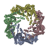
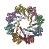
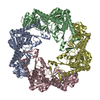
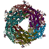
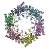
A: Signal peptide peptidase SppA
B: Signal peptide peptidase SppA
C: Signal peptide peptidase SppA
D: Signal peptide peptidase SppA
E: Signal peptide peptidase SppA
F: Signal peptide peptidase SppA
G: Signal peptide peptidase SppA
H: Signal peptide peptidase SppA


Theoretical massNumber of molelcules
Total (without water)240,0338
Polymers240,0338
Non-polymers00
Water3,981221
1


  • Idetical with deposited unit
  • defined by author&software
TypeNameSymmetry operationNumber
identity operation1_555x,y,z1
Buried area35370 Å2
ΔGint-202 kcal/mol
Surface area70500 Å2
MethodPISA
Unit cell
Length a, b, c (Å)87.797, 130.979, 207.099
Angle α, β, γ (deg.)90.00, 90.00, 90.00
Int Tables number19
Space group name H-MP212121

-
Components

#1: Protein
Signal peptide peptidase SppA


Mass: 30004.143 Da / Num. of mol.: 8 / Fragment: unp residues 51-335 / Mutation: K199A
Source method: isolated from a genetically manipulated source
Source: (gene. exp.) Bacillus subtilis subsp. subtilis (bacteria)
Strain: 168
Gene: BSU29530, CLONED SIGNAL PEPTIDE PEPTIDASE A GENE FROM RESIDUES 26 TO 335, LYS199 MUTATED TO ALA, sppA, yteI
Plasmid: PET28B / Production host: Escherichia coli (E. coli) / Strain (production host): TUNER(DE3)
References: UniProt: O34525, Hydrolases; Acting on peptide bonds (peptidases); Serine endopeptidases
#2: Water ChemComp-HOH / water / Water


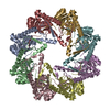
Mass: 18.015 Da / Num. of mol.: 221 / Source method: isolated from a natural source / Formula: H2O
Sequence detailsLIMITED PROTEOLYSIS WAS CARRIED OUT ON SPPA_BS DELTA 1-25 K199A USING THERMOLYSIN, FOLLOWED BY SIZE- ...LIMITED PROTEOLYSIS WAS CARRIED OUT ON SPPA_BS DELTA 1-25 K199A USING THERMOLYSIN, FOLLOWED BY SIZE-EXCLUSION CHROMATOGRAPHY. THE CRYSTALLIZED AND SOLVED STRUCTURE IS A PROTEASE RESISTANT FRAGMENT OF SPPA_BS DELTA 1-25 K199A. USING N-TERMINAL SEQUENCING AND POSITIVE ELECTROSPRAY IONIZATION MASS SPECTROMETRY, THE PROTEASE RESISTANT FRAGMENT WAS IDENTIFIED AS SPPA_BS 51-295. THE REFINED OCTAMERIC STRUCTURE REVEALED THE CORE DOMAIN OF SPPA_BS 51-295, WITH THE C-TERMINI OF SPPA_BS BOUND IN THE EIGHT ACTIVE SITES. THE IDENTITY OF THE PEPTIDE WAS CONFIRMED AS 308-335 OF SPPA_BS(FKSEIDFLNMREILSQSGSPRMMYLYAK) BY TANDEM MASS SPECTROMETRY FRAGMENTATION ANALYSIS. CLEAR ELECTRON DESITY IS OBSERVED FOR 326-335 (SPRMMYLYAK). WHAT HAS BEEN CRYSTALLIZED IS SPPA_BS RESIDUES: 51-295 & 308-335

-
Experimental details

-
Experiment

ExperimentMethod: X-RAY DIFFRACTION / Number of used crystals: 1

-
Sample preparation

CrystalDensity Matthews: 2.48 Å3/Da / Density % sol: 50.42 %
Crystal growTemperature: 298.15 K / Method: vapor diffusion, sitting drop / pH: 8.5
Details: 100mM TRIS, 23% TERT-BUTANOL UNDER PARAFFIN OIL, VAPOR DIFFUSION, SITTING DROP, temperature 298.15K, pH 8.5

-
Data collection

DiffractionMean temperature: 100 K
Diffraction sourceSource: SYNCHROTRON / Site: CLSI / Beamline: 08ID-1 / Wavelength: 0.97949 / Wavelength: 0.97949 Å
DetectorType: RAYONIX MX-300 / Detector: CCD / Date: Feb 25, 2011 / Details: VERTICAL COLLIMATING MIRROR
RadiationMonochromator: DOUBLE CRYSTAL MONOCHROMATOR (DCM) / Protocol: SINGLE WAVELENGTH / Monochromatic (M) / Laue (L): M / Scattering type: x-ray
Radiation wavelengthWavelength: 0.97949 Å / Relative weight: 1
ReflectionResolution: 2.39→50 Å / Num. all: 93912 / Num. obs: 93168 / % possible obs: 99.2 % / Observed criterion σ(F): 2 / Observed criterion σ(I): -3 / Redundancy: 6.8 % / Biso Wilson estimate: 51 Å2 / Rmerge(I) obs: 0.06 / Net I/σ(I): 50.3
Reflection shellResolution: 2.39→2.49 Å / Redundancy: 6.3 % / Rmerge(I) obs: 0.302 / Mean I/σ(I) obs: 6.7 / Num. unique all: 9085 / % possible all: 97.5

-
Processing

Software
NameVersionClassification
MxDCdata collection
PHASERFOR MR 2.1phasing
REFMAC5.5.0109refinement
HKL-2000data reduction
HKL-2000data scaling
RefinementMethod to determine structure: MOLECULAR REPLACEMENT
Starting model: 3BFO
Resolution: 2.39→50 Å / Cor.coef. Fo:Fc: 0.943 / Cor.coef. Fo:Fc free: 0.926 / SU B: 15.86 / SU ML: 0.17 / Cross valid method: THROUGHOUT / ESU R Free: 0.235 / Stereochemistry target values: MAXIMUM LIKELIHOOD
RfactorNum. reflection% reflectionSelection details
Rfree0.24 4708 5 %RANDOM
Rwork0.206 ---
all0.285 93310 --
obs0.208 88919 98.7 %-
Solvent computationIon probe radii: 0.8 Å / Shrinkage radii: 0.8 Å / VDW probe radii: 1.4 Å / Solvent model: MASK
Displacement parametersBiso mean: 50.9 Å2
Baniso -1Baniso -2Baniso -3
1--0.36 Å20 Å20 Å2
2--1.59 Å20 Å2
3----1.23 Å2
Refinement stepCycle: LAST / Resolution: 2.39→50 Å
ProteinNucleic acidLigandSolventTotal
Num. atoms14103 0 0 221 14324
Refine LS restraints
Refine-IDTypeDev idealDev ideal targetNumber
X-RAY DIFFRACTIONr_bond_refined_d0.0080.02214250
X-RAY DIFFRACTIONr_angle_refined_deg1.0611.97719135
X-RAY DIFFRACTIONr_dihedral_angle_1_deg5.25751841
X-RAY DIFFRACTIONr_dihedral_angle_2_deg35.13425.028539
X-RAY DIFFRACTIONr_dihedral_angle_3_deg15.578152644
X-RAY DIFFRACTIONr_dihedral_angle_4_deg14.9691551
X-RAY DIFFRACTIONr_chiral_restr0.0690.22179
X-RAY DIFFRACTIONr_gen_planes_refined0.0040.02110314
X-RAY DIFFRACTIONr_mcbond_it0.4511.59195
X-RAY DIFFRACTIONr_mcangle_it0.898214663
X-RAY DIFFRACTIONr_scbond_it1.52435055
X-RAY DIFFRACTIONr_scangle_it2.5764.54472
LS refinement shellResolution: 2.39→2.46 Å / Total num. of bins used: 20
RfactorNum. reflection% reflection
Rfree0.307 305 -
Rwork0.243 6133 -
obs--93.14 %
Refinement TLS params.

Method: refined / Refine-ID: X-RAY DIFFRACTION

IDL112)L122)L132)L222)L232)L332)S11 (Å °)S12 (Å °)S13 (Å °)S21 (Å °)S22 (Å °)S23 (Å °)S31 (Å °)S32 (Å °)S33 (Å °)T112)T122)T132)T222)T232)T332)Origin x (Å)Origin y (Å)Origin z (Å)
11.20120.51870.34491.7322-0.06321.39450.0203-0.0007-0.01750.1339-0.01340.02530.00120.0258-0.00690.01570.00270.01240.17610.04760.030612.585618.9395-23.4787
20.48880.21880.25811.93661.57953.84790.04290.06040.06310.0009-0.1920.2982-0.2908-0.25020.14920.0683-0.05290.01260.24350.03110.155421.560534.0636-1.1891
31.0275-0.1741-0.50421.29460.32922.632-0.02730.1091-0.1112-0.0208-0.12560.19950.0869-0.14140.15280.099-0.1023-0.0420.138-0.0070.106515.673433.36526.617
42.91591.7922-1.82042.6158-0.8792.41250.24130.17760.5050.2298-0.04320.4779-0.1351-0.1734-0.19810.2493-0.0760.03410.1624-0.02580.2120.764516.48244.2149
53.10272.10370.34983.131.12931.6684-0.12320.13910.5559-0.0868-0.12740.4927-0.0903-0.20290.25070.2157-0.02720.02710.1530.00550.1599-17.0332-5.80540.1278
62.4350.65831.30160.93860.7952.5356-0.08590.03730.3855-0.0128-0.03940.0691-0.20190.0110.12520.1053-0.03030.04670.02240.00760.1274-25.8887-21.14317.0644
71.00550.1195-0.15120.7907-0.62184.9629-0.00030.09550.16740.02410.08790.2142-0.3889-0.6097-0.08760.12190.0359-0.00680.14550.01780.1537-20.3456-21.3729-10.9603
81.12040.79530.27841.749-0.60081.6435-0.23850.09180.2287-0.1150.15570.2668-0.2492-0.04270.08280.17070.0266-0.05360.1125-0.00210.0987-4.6205-4.2085-27.6063
Refinement TLS group
IDRefine-IDRefine TLS-IDAuth asym-IDAuth seq-ID
1X-RAY DIFFRACTION1A56 - 335
2X-RAY DIFFRACTION2B58 - 335
3X-RAY DIFFRACTION3C57 - 334
4X-RAY DIFFRACTION4D59 - 333
5X-RAY DIFFRACTION5E57 - 334
6X-RAY DIFFRACTION6F57 - 334
7X-RAY DIFFRACTION7G57 - 334
8X-RAY DIFFRACTION8H58 - 334

+
About Yorodumi

-
News

-
Feb 9, 2022. New format data for meta-information of EMDB entries

New format data for meta-information of EMDB entries

  • Version 3 of the EMDB header file is now the official format.
  • The previous official version 1.9 will be removed from the archive.

Related info.:EMDB header

External links:wwPDB to switch to version 3 of the EMDB data model

-
Aug 12, 2020. Covid-19 info

Covid-19 info

URL: https://pdbj.org/emnavi/covid19.php

New page: Covid-19 featured information page in EM Navigator.

Related info.:Covid-19 info / Mar 5, 2020. Novel coronavirus structure data

+
Mar 5, 2020. Novel coronavirus structure data

Novel coronavirus structure data

Related info.:Yorodumi Speices / Aug 12, 2020. Covid-19 info

External links:COVID-19 featured content - PDBj / Molecule of the Month (242):Coronavirus Proteases

+
Jan 31, 2019. EMDB accession codes are about to change! (news from PDBe EMDB page)

EMDB accession codes are about to change! (news from PDBe EMDB page)

  • The allocation of 4 digits for EMDB accession codes will soon come to an end. Whilst these codes will remain in use, new EMDB accession codes will include an additional digit and will expand incrementally as the available range of codes is exhausted. The current 4-digit format prefixed with “EMD-” (i.e. EMD-XXXX) will advance to a 5-digit format (i.e. EMD-XXXXX), and so on. It is currently estimated that the 4-digit codes will be depleted around Spring 2019, at which point the 5-digit format will come into force.
  • The EM Navigator/Yorodumi systems omit the EMD- prefix.

Related info.:Q: What is EMD? / ID/Accession-code notation in Yorodumi/EM Navigator

External links:EMDB Accession Codes are Changing Soon! / Contact to PDBj

+
Jul 12, 2017. Major update of PDB

Major update of PDB

  • wwPDB released updated PDB data conforming to the new PDBx/mmCIF dictionary.
  • This is a major update changing the version number from 4 to 5, and with Remediation, in which all the entries are updated.
  • In this update, many items about electron microscopy experimental information are reorganized (e.g. em_software).
  • Now, EM Navigator and Yorodumi are based on the updated data.

External links:wwPDB Remediation / Enriched Model Files Conforming to OneDep Data Standards Now Available in the PDB FTP Archive

-
Yorodumi

Thousand views of thousand structures

  • Yorodumi is a browser for structure data from EMDB, PDB, SASBDB, etc.
  • This page is also the successor to EM Navigator detail page, and also detail information page/front-end page for Omokage search.
  • The word "yorodu" (or yorozu) is an old Japanese word meaning "ten thousand". "mi" (miru) is to see.

Related info.:EMDB / PDB / SASBDB / Comparison of 3 databanks / Yorodumi Search / Aug 31, 2016. New EM Navigator & Yorodumi / Yorodumi Papers / Jmol/JSmol / Function and homology information / Changes in new EM Navigator and Yorodumi

Read more