 Movie
Movie Controller


+ Open data

- Basic information
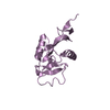
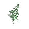
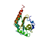
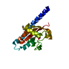
| Entry | Database: PDB / ID: 2f73 | ||||||
|---|---|---|---|---|---|---|---|
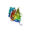
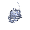
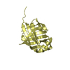
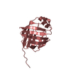
| Title | Crystal structure of human fatty acid binding protein 1 (FABP1) | ||||||
 Components Components | Fatty acid-binding protein, liver | ||||||
 Keywords Keywords | LIPID BINDING PROTEIN /  fatty acid binding protein / Structural Genomics / Structural Genomics Consortium / SGC fatty acid binding protein / Structural Genomics / Structural Genomics Consortium / SGC | ||||||
| Function / homology |  Function and homology information Function and homology informationresponse to vitamin B3 / oleic acid binding / apical cortex / positive regulation of fatty acid beta-oxidation / bile acid binding / intestinal absorption / Heme degradation / long-chain fatty acid transmembrane transporter activity / heterocyclic compound binding / Triglyceride catabolism ...response to vitamin B3 / oleic acid binding / apical cortex / positive regulation of fatty acid beta-oxidation / bile acid binding / intestinal absorption / Heme degradation / long-chain fatty acid transmembrane transporter activity / heterocyclic compound binding / Triglyceride catabolism / antioxidant activity / peroxisomal matrix / fatty acid transport / Regulation of lipid metabolism by PPARalpha / fatty acid binding / phospholipid binding / negative regulation of cysteine-type endopeptidase activity involved in apoptotic process / PPARA activates gene expression / Cytoprotection by HMOX1 / cellular response to hydrogen peroxide / cellular response to hypoxia / chromatin binding / negative regulation of apoptotic process / protein-containing complex / extracellular exosome / nucleoplasm / nucleus / cytosolSimilarity search - Function | ||||||
| Biological species |  Homo sapiens (human) Homo sapiens (human) | ||||||
| Method | X-RAY DIFFRACTION / SYNCHROTRON / MOLECULAR REPLACEMENT / Resolution: 2.5 Å | ||||||
 Authors Authors | Kursula, P. / Thorsell, A.G. / Arrowsmith, C. / Berglund, H. / Edwards, A. / Ehn, M. / Flodin, S. / Graslund, S. / Hammarstrom, M. / Holmberg Schiavone, L. ...Kursula, P. / Thorsell, A.G. / Arrowsmith, C. / Berglund, H. / Edwards, A. / Ehn, M. / Flodin, S. / Graslund, S. / Hammarstrom, M. / Holmberg Schiavone, L. / Kotenyova, T. / Nilsson-Ehle, P. / Nordlund, P. / Nyman, T. / Ogg, D. / Persson, C. / Sagemark, J. / Stenmark, P. / Sundstrom, M. / van den Berg, S. / Weigelt, J. / Hallberg, B.M. / Structural Genomics Consortium (SGC) | ||||||
 Citation Citation | Journal: To be Published Title: Crystal structure of human FABP1 Authors: Kursula, P. / Thorsell, A.G. / Arrowsmith, C. / Berglund, H. / Edwards, A. / Ehn, M. / Flodin, S. / Graslund, S. / Hammarstrom, M. / Holmberg Schiavone, L. / Kotenyova, T. / Nilsson-Ehle, P. ...Authors: Kursula, P. / Thorsell, A.G. / Arrowsmith, C. / Berglund, H. / Edwards, A. / Ehn, M. / Flodin, S. / Graslund, S. / Hammarstrom, M. / Holmberg Schiavone, L. / Kotenyova, T. / Nilsson-Ehle, P. / Nordlund, P. / Nyman, T. / Ogg, D. / Persson, C. / Sagemark, J. / Stenmark, P. / Sundstrom, M. / van den Berg, S. / Weigelt, J. / Hallberg, B.M. / Structural Genomics Consortium (SGC) | ||||||
| History |
|
- Structure visualization
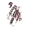
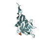
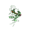
| Structure viewer | Molecule: MolmilJmol/JSmol |
|---|
- Downloads & links
-Download
| PDBx/mmCIF format | 2f73.cif.gz | 218.1 KB | Display | PDBx/mmCIF format |
|---|---|---|---|---|
| PDB format | pdb2f73.ent.gz | 176.8 KB | Display | PDB format |
| PDBx/mmJSON format | 2f73.json.gz | Tree view | PDBx/mmJSON format | |
| Others |  Other downloads Other downloads |
-Validation report
| Arichive directory | https://data.pdbj.org/pub/pdb/validation_reports/f7/2f73ftp://data.pdbj.org/pub/pdb/validation_reports/f7/2f73 | HTTPS FTP |
|---|
-Related structure data
| Related structure data | 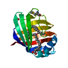 1lfoS S: Starting model for refinement |
|---|---|
| Similar structure data |


-Links
 PDBj
PDBj


- Assembly


-Components
| #1: Protein | Mass: 16785.117 Da / Num. of mol.: 8 Source method: isolated from a genetically manipulated source Source: (gene. exp.) Homo sapiens (human) / Gene: FABP1, FABPL / Production host:  Escherichia coli (E. coli) / References: UniProt: P07148 Escherichia coli (E. coli) / References: UniProt: P07148#2: Water | ChemComp-HOH / | Water Mass: 18.015 Da / Num. of mol.: 40 / Source method: isolated from a natural source / Formula: H2O Mass: 18.015 Da / Num. of mol.: 40 / Source method: isolated from a natural source / Formula: H2O |
|---|
-Experimental details
-Experiment
| Experiment | Method: X-RAY DIFFRACTION / Number of used crystals: 1 |
|---|
- Sample preparation
| Crystal | Density Matthews: 2.6 Å3/Da / Density % sol: 52.71 % |
|---|---|
| Crystal grow | Method: vapor diffusion / Details: VAPOR DIFFUSION |
-Data collection
| Diffraction | Mean temperature: 133 K |
|---|---|
| Diffraction source | Source: SYNCHROTRON / Site: BESSY  / Beamline: 14.1 / Wavelength: 0.9781 Å / Beamline: 14.1 / Wavelength: 0.9781 Å |
| Detector | Type: MARRESEARCH / Detector: CCD / Date: Nov 22, 2005 |
| Radiation | Protocol: SINGLE WAVELENGTH / Monochromatic (M) / Laue (L): M / Scattering type: x-ray |
| Radiation wavelength | Wavelength: 0.9781 Å / Relative weight: 1 |
| Reflection | Resolution: 2.5→20 Å / Num. all: 49103 / Num. obs: 49103 / % possible obs: 99.4 % / Observed criterion σ(F): -3 / Observed criterion σ(I): -3 / Redundancy: 4.3 % / Biso Wilson estimate: 50 Å2 / Rsym value: 0.084 / Net I/σ(I): 13.2 |
| Reflection shell | Resolution: 2.5→2.6 Å / Redundancy: 4.4 % / Mean I/σ(I) obs: 2.6 / Num. unique all: 5391 / Rsym value: 0.655 / % possible all: 99.8 |
- Processing
| Software |
| |||||||||||||||||||||||||
|---|---|---|---|---|---|---|---|---|---|---|---|---|---|---|---|---|---|---|---|---|---|---|---|---|---|---|
| Refinement | Method to determine structure: MOLECULAR REPLACEMENT Starting model: 1LFO Resolution: 2.5→20 Å / σ(F): -3 / Stereochemistry target values: Engh & Huber Details: The twinning operator k,h,-l and the twinning fraction 0.27 were used in the refinement. THis is pseudomerohedral twinning.
| |||||||||||||||||||||||||
| Refinement step | Cycle: LAST / Resolution: 2.5→20 Å
| |||||||||||||||||||||||||
| Refine LS restraints |
|
