- PDB-1mai: STRUCTURE OF THE PLECKSTRIN HOMOLOGY DOMAIN FROM PHOSPHOLIPASE C ... -
+
Open data
ID or keywords:
Loading...
-
Basic information
Entry
Database: PDB / ID: 1mai
Title
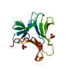
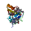
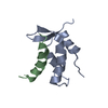
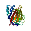
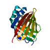
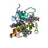
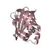
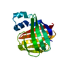
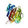
STRUCTURE OF THE PLECKSTRIN HOMOLOGY DOMAIN FROM PHOSPHOLIPASE C DELTA IN COMPLEX WITH INOSITOL TRISPHOSPHATE
Components
PHOSPHOLIPASE C DELTA-1
Keywords
SIGNAL TRANSDUCTION PROTEIN / PLECKSTRIN / PHOSPHOLIPASE / INOSITOL TRISPHOSPHATE / HYDROLASE
Function / homology
Function and homology information
Synthesis of IP3 and IP4 in the cytosol / response to prostaglandin F / positive regulation of inositol trisphosphate biosynthetic process / phosphatidylinositol phosphate binding / phosphoinositide phospholipase C / positive regulation of norepinephrine secretion / phosphatidylinositol metabolic process / response to aluminum ion / phosphatidylinositol phospholipase C activity / phosphatidic acid binding ...Synthesis of IP3 and IP4 in the cytosol / response to prostaglandin F / positive regulation of inositol trisphosphate biosynthetic process / phosphatidylinositol phosphate binding / phosphoinositide phospholipase C / positive regulation of norepinephrine secretion / phosphatidylinositol metabolic process / response to aluminum ion / phosphatidylinositol phospholipase C activity / phosphatidic acid binding / phospholipase C activity / inositol 1,4,5 trisphosphate binding / calcium-dependent phospholipid binding / phosphatidylinositol-mediated signaling / GTPase activating protein binding / labyrinthine layer blood vessel development / response to hyperoxia / lipid catabolic process / release of sequestered calcium ion into cytosol / response to organonitrogen compound / regulation of cytosolic calcium ion concentration / phosphatidylinositol-4,5-bisphosphate binding / mitochondrial membrane / phospholipid binding / response to peptide hormone / response to calcium ion / phospholipase C-activating G protein-coupled receptor signaling pathway / regulation of cell population proliferation / angiogenesis / G protein-coupled receptor signaling pathway / calcium ion binding / enzyme binding / cytosol / cytoplasm Similarity search - Function
In the structure databanks used in Yorodumi, some data are registered as the other names, "COVID-19 virus" and "2019-nCoV". Here are the details of the virus and the list of structure data.
Jan 31, 2019. EMDB accession codes are about to change! (news from PDBe EMDB page)
EMDB accession codes are about to change! (news from PDBe EMDB page)
The allocation of 4 digits for EMDB accession codes will soon come to an end. Whilst these codes will remain in use, new EMDB accession codes will include an additional digit and will expand incrementally as the available range of codes is exhausted. The current 4-digit format prefixed with “EMD-” (i.e. EMD-XXXX) will advance to a 5-digit format (i.e. EMD-XXXXX), and so on. It is currently estimated that the 4-digit codes will be depleted around Spring 2019, at which point the 5-digit format will come into force.
The EM Navigator/Yorodumi systems omit the EMD- prefix.
Related info.:Q: What is EMD? / ID/Accession-code notation in Yorodumi/EM Navigator
Yorodumi is a browser for structure data from EMDB, PDB, SASBDB, etc.
This page is also the successor to EM Navigator detail page, and also detail information page/front-end page for Omokage search.
The word "yorodu" (or yorozu) is an old Japanese word meaning "ten thousand". "mi" (miru) is to see.
Related info.:EMDB / PDB / SASBDB / Comparison of 3 databanks / Yorodumi Search / Aug 31, 2016. New EM Navigator & Yorodumi / Yorodumi Papers / Jmol/JSmol / Function and homology information / Changes in new EM Navigator and Yorodumi
 Movie
Movie Controller

 Yorodumi
 Open data
 Basic information
 Components
 Keywords
 SIGNAL TRANSDUCTION PROTEIN /
  Function and homology information
 Authors
 Citation
 Structure visualization
 Downloads & links
 Other downloads
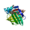
 PDBj
PDBj


 Assembly
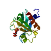

 Mass: 420.096 Da / Num. of mol.: 1 / Source method: obtained synthetically / Formula: C6H15O15P3
 Mass: 18.015 Da / Num. of mol.: 110 / Source method: isolated from a natural source / Formula: H2O
 Sample preparation
 / Beamline: X25 / Wavelength: 1
  Processing