 Movie
Movie Controller


+ Open data

- Basic information
| Entry | Database: PDB / ID: 5ljc | ||||||
|---|---|---|---|---|---|---|---|
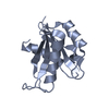
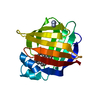
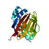
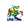
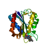
| Title | Crystal structure of holo human CRBP1 | ||||||
 Components Components | Retinol-binding protein 1 | ||||||
 Keywords Keywords | retinol-binding protein / Retinol / Vitamin A / RBP | ||||||
| Function / homology |  Function and homology information Function and homology informationDefective visual phototransduction due to LRAT loss of function / all-trans-retinol binding / retinoic acid biosynthetic process / vitamin A metabolic process / retinoid binding / retinal binding / The canonical retinoid cycle in rods (twilight vision) / lipid homeostasis / fatty acid transport / Retinoid metabolism and transport ...Defective visual phototransduction due to LRAT loss of function / all-trans-retinol binding / retinoic acid biosynthetic process / vitamin A metabolic process / retinoid binding / retinal binding / The canonical retinoid cycle in rods (twilight vision) / lipid homeostasis / fatty acid transport / Retinoid metabolism and transport / lipid droplet / fatty acid binding / nucleoplasm / nucleus / cytosol Similarity search - Function | ||||||
| Biological species |  Homo sapiens (human) Homo sapiens (human) | ||||||
| Method |  X-RAY DIFFRACTION / SYNCHROTRON / MOLECULAR REPLACEMENT / Resolution: 1.433 Å X-RAY DIFFRACTION / SYNCHROTRON / MOLECULAR REPLACEMENT / Resolution: 1.433 Å | ||||||
 Authors Authors | Zanotti, G. / Vallese, F. / Berni, R. / Menozzi, I. | ||||||
 Citation Citation | Journal: J. Struct. Biol. / Year: 2017 Title: Structural and molecular determinants affecting the interaction of retinol with human CRBP1. Authors: Menozzi, I. / Vallese, F. / Polverini, E. / Folli, C. / Berni, R. / Zanotti, G. | ||||||
| History |
|
- Structure visualization
| Structure viewer | Molecule: MolmilJmol/JSmol |
|---|
- Downloads & links
-Download
| PDBx/mmCIF format | 5ljc.cif.gz | 73.1 KB | Display | PDBx/mmCIF format |
|---|---|---|---|---|
| PDB format | pdb5ljc.ent.gz | 54.2 KB | Display | PDB format |
| PDBx/mmJSON format | 5ljc.json.gz | Tree view | PDBx/mmJSON format | |
| Others |  Other downloads Other downloads |
-Validation report
| Arichive directory | https://data.pdbj.org/pub/pdb/validation_reports/lj/5ljcftp://data.pdbj.org/pub/pdb/validation_reports/lj/5ljc | HTTPS FTP |
|---|
-Related structure data
| Related structure data |  5ljbSC  5ljdC  5ljeC  5ljgC  5ljhC  5ljkC S: Starting model for refinement C: citing same article ( |
|---|---|
| Similar structure data |


-Links
 PDBj
PDBj


- Assembly
| Deposited unit | 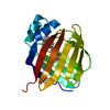
| ||||||||
|---|---|---|---|---|---|---|---|---|---|
| 1 |
| ||||||||
| Unit cell |
|
-Components
| #1: Protein | Mass: 15856.213 Da / Num. of mol.: 1 Source method: isolated from a genetically manipulated source Details: Position 108 was mutated in Leu. Q108L / Source: (gene. exp.) Homo sapiens (human) / Gene: RBP1, CRBP1 / Production host:  |
|---|---|
| #2: Chemical | ChemComp-RTL /   Mass: 286.452 Da / Num. of mol.: 1 / Source method: obtained synthetically / Formula: C20H30O Mass: 286.452 Da / Num. of mol.: 1 / Source method: obtained synthetically / Formula: C20H30O |
| #3: Chemical | ChemComp-NA /   Mass: 22.990 Da / Num. of mol.: 1 / Source method: obtained synthetically / Formula: Na Mass: 22.990 Da / Num. of mol.: 1 / Source method: obtained synthetically / Formula: Na |
| #4: Water | ChemComp-HOH /  Mass: 18.015 Da / Num. of mol.: 178 / Source method: isolated from a natural source / Formula: H2O Mass: 18.015 Da / Num. of mol.: 178 / Source method: isolated from a natural source / Formula: H2O |
-Experimental details
-Experiment
| Experiment | Method: X-RAY DIFFRACTION / Number of used crystals: 1 |
|---|
- Sample preparation
| Crystal | Density Matthews: 2.02 Å3/Da / Density % sol: 40 % |
|---|---|
| Crystal grow | Temperature: 279 K / Method: vapor diffusion / pH: 5 Details: 0.2 M ammonium chloride, 0.1 M sodium acetate, 0.2 mM zinc chloride, pH 5,0, 20% PEG 6000 |
-Data collection
| Diffraction | Mean temperature: 100 K |
|---|---|
| Diffraction source | Source: SYNCHROTRON / Site: ESRF  / Beamline: ID29 / Wavelength: 0.91881 Å / Beamline: ID29 / Wavelength: 0.91881 Å |
| Detector | Type: DECTRIS PILATUS 6M / Detector: PIXEL / Date: Mar 2, 2015 |
| Radiation | Protocol: SINGLE WAVELENGTH / Monochromatic (M) / Laue (L): M / Scattering type: x-ray |
| Radiation wavelength | Wavelength: 0.91881 Å / Relative weight: 1 |
| Reflection | Resolution: 1.43→40.883 Å / Num. obs: 23562 / % possible obs: 94.4 % / Observed criterion σ(F): 0 / Observed criterion σ(I): 0 / Redundancy: 3.3 % / Rmerge(I) obs: 0.063 / Net I/σ(I): 9.2 |
| Reflection shell | Resolution: 1.43→1.51 Å / Redundancy: 3 % / Rmerge(I) obs: 0.504 / % possible all: 82.5 |
- Processing
| Software |
| |||||||||||||||||||||||||||||||||||||||||||||||||||||||||||||||||||||||||||||||||||||||||||||||||||||||||||||||||||||||
|---|---|---|---|---|---|---|---|---|---|---|---|---|---|---|---|---|---|---|---|---|---|---|---|---|---|---|---|---|---|---|---|---|---|---|---|---|---|---|---|---|---|---|---|---|---|---|---|---|---|---|---|---|---|---|---|---|---|---|---|---|---|---|---|---|---|---|---|---|---|---|---|---|---|---|---|---|---|---|---|---|---|---|---|---|---|---|---|---|---|---|---|---|---|---|---|---|---|---|---|---|---|---|---|---|---|---|---|---|---|---|---|---|---|---|---|---|---|---|---|---|
| Refinement | Method to determine structure: MOLECULAR REPLACEMENT Starting model: 5LJB Resolution: 1.433→40.883 Å / SU ML: 0.18 / Cross valid method: FREE R-VALUE / σ(F): 1.25 / Phase error: 23.13
| |||||||||||||||||||||||||||||||||||||||||||||||||||||||||||||||||||||||||||||||||||||||||||||||||||||||||||||||||||||||
| Solvent computation | Shrinkage radii: 0.9 Å / VDW probe radii: 1.11 Å | |||||||||||||||||||||||||||||||||||||||||||||||||||||||||||||||||||||||||||||||||||||||||||||||||||||||||||||||||||||||
| Refinement step | Cycle: LAST / Resolution: 1.433→40.883 Å
| |||||||||||||||||||||||||||||||||||||||||||||||||||||||||||||||||||||||||||||||||||||||||||||||||||||||||||||||||||||||
| Refine LS restraints |
| |||||||||||||||||||||||||||||||||||||||||||||||||||||||||||||||||||||||||||||||||||||||||||||||||||||||||||||||||||||||
| LS refinement shell |
|
