 Movie
Movie Controller


+ Open data

- Basic information
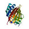
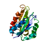
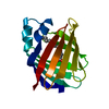
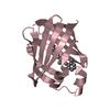
| Entry | Database: PDB / ID: 1p6p | ||||||
|---|---|---|---|---|---|---|---|
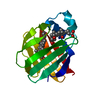
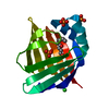
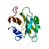
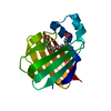
| Title | Crystal Structure of Toad Liver Basic Fatty Acid-Binding Protein | ||||||
 Components Components | Fatty acid-binding protein, liver | ||||||
 Keywords Keywords | LIPID BINDING PROTEIN / BETA BARREL | ||||||
| Function / homology |  Function and homology information Function and homology information | ||||||
| Biological species | Bufo arenarum (Argentine toad) | ||||||
| Method |  X-RAY DIFFRACTION / MOLECULAR REPLACEMENT / Resolution: 2.5 Å X-RAY DIFFRACTION / MOLECULAR REPLACEMENT / Resolution: 2.5 Å | ||||||
 Authors Authors | Di Pietro, S.M. / Corsico, B. / Perduca, M. / Monaco, H.L. / Santome, J.A. | ||||||
 Citation Citation | Journal: Biochemistry / Year: 2003 Title: Structural and Biochemical Characterization of Toad Liver Basic Fatty Acid-Binding Protein Authors: Di Pietro, S.M. / Corsico, B. / Perduca, M. / Monaco, H.L. / Santome, J.A. #1: Journal: Acta Crystallogr.,Sect.D / Year: 2001Title: Crystallization and Preliminary X-ray Study of two Liver Basic Fatty Acid-Binding Proteins Authors: Di Pietro, S.M. / Perduca, M. / Santome, J.A. / Monaco, H.L. | ||||||
| History |
|
- Structure visualization
| Structure viewer | Molecule: MolmilJmol/JSmol |
|---|
- Downloads & links
-Download
| PDBx/mmCIF format | 1p6p.cif.gz | 35.6 KB | Display | PDBx/mmCIF format |
|---|---|---|---|---|
| PDB format | pdb1p6p.ent.gz | 24.6 KB | Display | PDB format |
| PDBx/mmJSON format | 1p6p.json.gz | Tree view | PDBx/mmJSON format | |
| Others |  Other downloads Other downloads |
-Validation report
| Arichive directory | https://data.pdbj.org/pub/pdb/validation_reports/p6/1p6pftp://data.pdbj.org/pub/pdb/validation_reports/p6/1p6p | HTTPS FTP |
|---|
-Related structure data
| Similar structure data |
|---|


-Links
 PDBj
PDBj

- Assembly
| Deposited unit | 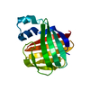
| ||||||||
|---|---|---|---|---|---|---|---|---|---|
| 1 |
| ||||||||
| Unit cell |
|
-Components
| #1: Protein | Mass: 13967.776 Da / Num. of mol.: 1 / Source method: isolated from a natural source / Source: (natural) Bufo arenarum (Argentine toad) / Organ: Liver / References: UniProt: P83409 |
|---|---|
| #2: Water | ChemComp-HOH /  Mass: 18.015 Da / Num. of mol.: 25 / Source method: isolated from a natural source / Formula: H2O Mass: 18.015 Da / Num. of mol.: 25 / Source method: isolated from a natural source / Formula: H2O |
-Experimental details
-Experiment
| Experiment | Method: X-RAY DIFFRACTION / Number of used crystals: 1 |
|---|
- Sample preparation
| Crystal | Density Matthews: 2.8 Å3/Da / Density % sol: 56.13 % | ||||||||||||||||||||||||||||||
|---|---|---|---|---|---|---|---|---|---|---|---|---|---|---|---|---|---|---|---|---|---|---|---|---|---|---|---|---|---|---|---|
| Crystal grow | Temperature: 277 K / Method: vapor diffusion, sitting drop / pH: 7.4 Details: Tris, PEG 1500, pH 7.4, VAPOR DIFFUSION, SITTING DROP, temperature 277.0K | ||||||||||||||||||||||||||||||
| Crystal grow | *PLUS Temperature: 277 K / Method: vapor diffusion, sitting dropDetails: Di Pietro, S.M., (2001) Acta Crystallogr.,Sect.D, 57, 1903. | ||||||||||||||||||||||||||||||
| Components of the solutions | *PLUS
|
-Data collection
| Diffraction | Mean temperature: 298 K |
|---|---|
| Diffraction source | Source: ROTATING ANODE / Type: RIGAKU RU200 / Wavelength: 1.5418 Å |
| Detector | Type: RIGAKU RAXIS IIC / Detector: IMAGE PLATE / Date: Dec 2, 1999 |
| Radiation | Monochromator: graphite / Protocol: SINGLE WAVELENGTH / Monochromatic (M) / Laue (L): M / Scattering type: x-ray |
| Radiation wavelength | Wavelength: 1.5418 Å / Relative weight: 1 |
| Reflection | Resolution: 2.5→20 Å / Num. all: 5917 / Num. obs: 5917 / % possible obs: 98.9 % / Observed criterion σ(F): 0 / Observed criterion σ(I): 0 / Redundancy: 8 % / Biso Wilson estimate: 37.7 Å2 / Rmerge(I) obs: 0.068 / Rsym value: 0.064 / Net I/σ(I): 11.5 |
| Reflection shell | Resolution: 2.5→2.64 Å / Redundancy: 9 % / Rmerge(I) obs: 0.267 / Mean I/σ(I) obs: 3 / Num. unique all: 832 / Rsym value: 0.252 / % possible all: 98.9 |
| Reflection | *PLUS Num. measured all: 47279 / Rmerge(I) obs: 0.064 |
| Reflection shell | *PLUS Highest resolution: 2.5 Å / % possible obs: 98.9 % / Rmerge(I) obs: 0.252 |
- Processing
| Software |
| |||||||||||||||||||||||||
|---|---|---|---|---|---|---|---|---|---|---|---|---|---|---|---|---|---|---|---|---|---|---|---|---|---|---|
| Refinement | Method to determine structure: MOLECULAR REPLACEMENT Starting model: axolotl liver basic FABP model (unpublished) Resolution: 2.5→20 Å / σ(F): 0 / σ(I): 0 / Stereochemistry target values: Engh & Huber
| |||||||||||||||||||||||||
| Refinement step | Cycle: LAST / Resolution: 2.5→20 Å
| |||||||||||||||||||||||||
| Refine LS restraints |
| |||||||||||||||||||||||||
| Refinement | *PLUS Num. reflection all: 5827 / Num. reflection obs: 5266 / Num. reflection Rfree: 561 | |||||||||||||||||||||||||
| Solvent computation | *PLUS | |||||||||||||||||||||||||
| Displacement parameters | *PLUS | |||||||||||||||||||||||||
| Refine LS restraints | *PLUS
| |||||||||||||||||||||||||
| LS refinement shell | *PLUS Highest resolution: 2.5 Å / Lowest resolution: 2.64 Å / Rfactor Rfree: 0.31 / Num. reflection Rfree: 54 / Rfactor Rwork: 0.269 / Num. reflection Rwork: 511 |
