 Movie
Movie Controller

[English] 日本語
 Yorodumi
- PDB-2ftb: Crystal structure of axolotl (Ambystoma mexicanum) liver bile aci... -
+ Open data

- Basic information
| Entry | Database: PDB / ID: 2ftb | ||||||
|---|---|---|---|---|---|---|---|
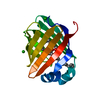
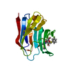
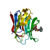
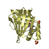
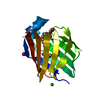
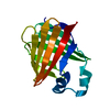
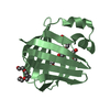
| Title | Crystal structure of axolotl (Ambystoma mexicanum) liver bile acid-binding protein bound to oleic acid | ||||||
 Components Components | Fatty acid-binding protein 2, liver | ||||||
 Keywords Keywords | LIPID BINDING PROTEIN / liver bile acid-binding protein / liver basic fatty acid-binding protein / axolotl / oleic acid / BABP / Lb-FABP | ||||||
| Function / homology |  Function and homology information Function and homology information | ||||||
| Biological species | Ambystoma mexicanum (axolotl) | ||||||
| Method |  X-RAY DIFFRACTION / MOLECULAR REPLACEMENT / Resolution: 2 Å X-RAY DIFFRACTION / MOLECULAR REPLACEMENT / Resolution: 2 Å | ||||||
 Authors Authors | Capaldi, S. / Guariento, M. / Perduca, M. / Di Pietro, S.M. / Santome, J.A. / Monaco, H.L. | ||||||
 Citation Citation | Journal: Proteins / Year: 2006 Title: Crystal structure of axolotl (Ambystoma mexicanum) liver bile acid-binding protein bound to cholic and oleic acid Authors: Capaldi, S. / Guariento, M. / Perduca, M. / Di Pietro, S.M. / Santome, J.A. / Monaco, H.L. #1: Journal: Acta Crystallogr.,Sect.D / Year: 2001 Title: Crystallization and preliminary X-ray study of two liver basic fatty acid-binding proteins Authors: Di Pietro, S.M. / Perduca, M. / Santome, J.A. / Monaco, H.L. | ||||||
| History |
|
- Structure visualization
| Structure viewer | Molecule: MolmilJmol/JSmol |
|---|
- Downloads & links
-Download
| PDBx/mmCIF format | 2ftb.cif.gz | 37.9 KB | Display | PDBx/mmCIF format |
|---|---|---|---|---|
| PDB format | pdb2ftb.ent.gz | 25.7 KB | Display | PDB format |
| PDBx/mmJSON format | 2ftb.json.gz | Tree view | PDBx/mmJSON format | |
| Others |  Other downloads Other downloads |
-Validation report
| Arichive directory | https://data.pdbj.org/pub/pdb/validation_reports/ft/2ftbftp://data.pdbj.org/pub/pdb/validation_reports/ft/2ftb | HTTPS FTP |
|---|
-Related structure data
| Related structure data |  2ft9C  1tvqS C: citing same article ( S: Starting model for refinement |
|---|---|
| Similar structure data |


-Links
 PDBj
PDBj


- Assembly
| Deposited unit | 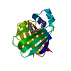
| ||||||||
|---|---|---|---|---|---|---|---|---|---|
| 1 |
| ||||||||
| Unit cell |
| ||||||||
| Components on special symmetry positions |
|
-Components
| #1: Protein | Mass: 13758.582 Da / Num. of mol.: 1 / Source method: isolated from a natural source / Source: (natural) Ambystoma mexicanum (axolotl) / Tissue: liver / References: UniProt: P81400 |
|---|---|
| #2: Chemical | ChemComp-OLA /   Mass: 282.461 Da / Num. of mol.: 1 / Source method: obtained synthetically / Formula: C18H34O2 Mass: 282.461 Da / Num. of mol.: 1 / Source method: obtained synthetically / Formula: C18H34O2 |
| #3: Water | ChemComp-HOH /  Mass: 18.015 Da / Num. of mol.: 37 / Source method: isolated from a natural source / Formula: H2O Mass: 18.015 Da / Num. of mol.: 37 / Source method: isolated from a natural source / Formula: H2O |
-Experimental details
-Experiment
| Experiment | Method: X-RAY DIFFRACTION / Number of used crystals: 1 |
|---|
- Sample preparation
| Crystal | Density Matthews: 2.47 Å3/Da / Density % sol: 50.29 % |
|---|---|
| Crystal grow | Temperature: 277 K / Method: vapor diffusion, hanging drop / pH: 8.5 Details: 30% PEG 4000, 0.2M sodium acetate, 0.1M Tris, pH 8.5, VAPOR DIFFUSION, HANGING DROP, temperature 277K |
-Data collection
| Diffraction | Mean temperature: 293 K |
|---|---|
| Diffraction source | Source: ROTATING ANODE / Type: RIGAKU RU200 / Wavelength: 1.5418 Å |
| Detector | Type: RIGAKU RAXIS II / Detector: IMAGE PLATE / Date: Feb 9, 2000 / Details: graphite crystal monochromator |
| Radiation | Monochromator: GRAPHITE / Protocol: SINGLE WAVELENGTH / Monochromatic (M) / Laue (L): M / Scattering type: x-ray |
| Radiation wavelength | Wavelength: 1.5418 Å / Relative weight: 1 |
| Reflection | Resolution: 2→45 Å / Num. all: 9823 / Num. obs: 9823 / % possible obs: 99.9 % / Observed criterion σ(F): 0 / Observed criterion σ(I): 0 / Rsym value: 0.041 |
| Reflection shell | Resolution: 2→2.05 Å / Rsym value: 0.185 / % possible all: 100 |
- Processing
| Software |
| ||||||||||||||||||||||||||||||||||||||||||||||||||||||||||||||||||||||||||||||||||||||||||
|---|---|---|---|---|---|---|---|---|---|---|---|---|---|---|---|---|---|---|---|---|---|---|---|---|---|---|---|---|---|---|---|---|---|---|---|---|---|---|---|---|---|---|---|---|---|---|---|---|---|---|---|---|---|---|---|---|---|---|---|---|---|---|---|---|---|---|---|---|---|---|---|---|---|---|---|---|---|---|---|---|---|---|---|---|---|---|---|---|---|---|---|
| Refinement | Method to determine structure: MOLECULAR REPLACEMENT Starting model: PDB ENTRY 1TVQ Resolution: 2→20 Å / Cor.coef. Fo:Fc: 0.938 / Cor.coef. Fo:Fc free: 0.902 / SU B: 3.95 / SU ML: 0.114 / Cross valid method: THROUGHOUT / σ(F): 0 / ESU R: 0.194 / ESU R Free: 0.177 / Stereochemistry target values: MAXIMUM LIKELIHOOD / Details: HYDROGENS HAVE BEEN ADDED IN THE RIDING POSITIONS
| ||||||||||||||||||||||||||||||||||||||||||||||||||||||||||||||||||||||||||||||||||||||||||
| Solvent computation | Ion probe radii: 0.8 Å / Shrinkage radii: 0.8 Å / VDW probe radii: 1.2 Å / Solvent model: MASK | ||||||||||||||||||||||||||||||||||||||||||||||||||||||||||||||||||||||||||||||||||||||||||
| Displacement parameters | Biso mean: 23.801 Å2
| ||||||||||||||||||||||||||||||||||||||||||||||||||||||||||||||||||||||||||||||||||||||||||
| Refinement step | Cycle: LAST / Resolution: 2→20 Å
| ||||||||||||||||||||||||||||||||||||||||||||||||||||||||||||||||||||||||||||||||||||||||||
| Refine LS restraints |
| ||||||||||||||||||||||||||||||||||||||||||||||||||||||||||||||||||||||||||||||||||||||||||
| LS refinement shell | Resolution: 2→2.052 Å / Total num. of bins used: 20
|
