 Movie
Movie Controller

[English] 日本語
 Yorodumi
- PDB-1f5w: DIMERIC STRUCTURE OF THE COXSACKIE VIRUS AND ADENOVIRUS RECEPTOR ... -
+ Open data

- Basic information
| Entry | Database: PDB / ID: 1f5w | ||||||
|---|---|---|---|---|---|---|---|
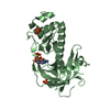
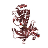
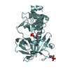
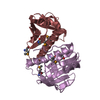
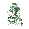
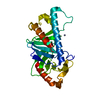
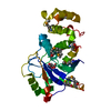
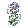
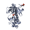
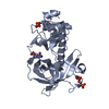
| Title | DIMERIC STRUCTURE OF THE COXSACKIE VIRUS AND ADENOVIRUS RECEPTOR D1 DOMAIN | ||||||
 Components Components | COXSACKIE VIRUS AND ADENOVIRUS RECEPTOR | ||||||
 Keywords Keywords |  Viral protein receptor / immunoglobulin V domain fold / symmetric dimer Viral protein receptor / immunoglobulin V domain fold / symmetric dimer | ||||||
| Function / homology |  Function and homology information Function and homology informationAV node cell-bundle of His cell adhesion involved in cell communication / cell adhesive protein binding involved in AV node cell-bundle of His cell communication / homotypic cell-cell adhesion / AV node cell to bundle of His cell communication / epithelial structure maintenance / gamma-delta T cell activation / regulation of AV node cell action potential / germ cell migration / apicolateral plasma membrane / cell-cell junction organization ...AV node cell-bundle of His cell adhesion involved in cell communication / cell adhesive protein binding involved in AV node cell-bundle of His cell communication / homotypic cell-cell adhesion / AV node cell to bundle of His cell communication / epithelial structure maintenance / gamma-delta T cell activation / regulation of AV node cell action potential / germ cell migration / apicolateral plasma membrane / cell-cell junction organization / transepithelial transport / connexin binding / cardiac muscle cell development / heterophilic cell-cell adhesion via plasma membrane cell adhesion molecules / bicellular tight junction / intercalated disc / cell adhesion molecule binding / mitochondrion organization / neutrophil chemotaxis / acrosomal vesicle / filopodium / PDZ domain binding / Cell surface interactions at the vascular wall / adherens junction / neuromuscular junction / beta-catenin binding / Immunoregulatory interactions between a Lymphoid and a non-Lymphoid cell / cell-cell junction / integrin binding / virus receptor activity / cell junction / cell body / heart development / growth cone / actin cytoskeleton organization / basolateral plasma membrane / defense response to virus / neuron projection / membrane raft / signaling receptor binding / protein-containing complex / extracellular space / extracellular region / nucleoplasm / identical protein binding / plasma membrane / cytoplasmSimilarity search - Function | ||||||
| Biological species |  Homo sapiens (human) Homo sapiens (human) | ||||||
| Method | X-RAY DIFFRACTION / SYNCHROTRON / MOLECULAR REPLACEMENT / Resolution: 1.7 Å | ||||||
 Authors Authors | van Raaij, M.J. / Chouin, E. / van der Zandt, H. / Bergelson, J.M. / Cusack, S. | ||||||
 Citation Citation | Journal: Structure Fold.Des. / Year: 2000 Title: Dimeric structure of the coxsackievirus and adenovirus receptor D1 domain at 1.7 A resolution. Authors: van Raaij, M.J. / Chouin, E. / van der Zandt, H. / Bergelson, J.M. / Cusack, S. | ||||||
| History |
|
- Structure visualization
| Structure viewer | Molecule: MolmilJmol/JSmol |
|---|
- Downloads & links
-Download
| PDBx/mmCIF format | 1f5w.cif.gz | 117.1 KB | Display | PDBx/mmCIF format |
|---|---|---|---|---|
| PDB format | pdb1f5w.ent.gz | 91.4 KB | Display | PDB format |
| PDBx/mmJSON format | 1f5w.json.gz | Tree view | PDBx/mmJSON format | |
| Others |  Other downloads Other downloads |
-Validation report
| Arichive directory | https://data.pdbj.org/pub/pdb/validation_reports/f5/1f5wftp://data.pdbj.org/pub/pdb/validation_reports/f5/1f5w | HTTPS FTP |
|---|
-Related structure data
| Related structure data | 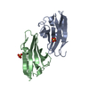 1eajC 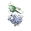 1kacS S: Starting model for refinement C: citing same article ( |
|---|---|
| Similar structure data |
-Links
 PDBj
PDBj


- Assembly
| Deposited unit | 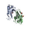
| ||||||||
|---|---|---|---|---|---|---|---|---|---|
| 1 |
| ||||||||
| 2 | 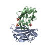
| ||||||||
| Unit cell |
| ||||||||
| Details | The biological assembly is the dimer formed by chains A and B |
-Components
| #1: Protein | Mass: 14048.972 Da / Num. of mol.: 2 / Fragment: D1 DOMAIN Source method: isolated from a genetically manipulated source Source: (gene. exp.) Homo sapiens (human) / Description: HELA CELL CDNA LIBRARY / Plasmid: PAB3 / Production host:  Escherichia coli (E. coli) / References: UniProt: P78310 Escherichia coli (E. coli) / References: UniProt: P78310#2: Chemical | Sulfate  Mass: 96.063 Da / Num. of mol.: 3 / Source method: obtained synthetically / Formula: SO4 Mass: 96.063 Da / Num. of mol.: 3 / Source method: obtained synthetically / Formula: SO4#3: Water | ChemComp-HOH / | Water Mass: 18.015 Da / Num. of mol.: 232 / Source method: isolated from a natural source / Formula: H2O Mass: 18.015 Da / Num. of mol.: 232 / Source method: isolated from a natural source / Formula: H2O |
|---|
-Experimental details
-Experiment
| Experiment | Method: X-RAY DIFFRACTION / Number of used crystals: 1 |
|---|
- Sample preparation
| Crystal | Density Matthews: 3.04 Å3/Da / Density % sol: 59.3 % Description: THE SIGMA(I) IN SCALA APPEARS TO BE OVERESTIMATED FOR OUR DATASET, GIVING LOW I/SIGMA(I) VALUES. THE Mn(I)/sd OVERALL AND FOR THE HIGH RESOLUTION SHELL ARE 10.8 AND 3.2, RESPECTIVELY TWO ...Description: THE SIGMA(I) IN SCALA APPEARS TO BE OVERESTIMATED FOR OUR DATASET, GIVING LOW I/SIGMA(I) VALUES. THE Mn(I)/sd OVERALL AND FOR THE HIGH RESOLUTION SHELL ARE 10.8 AND 3.2, RESPECTIVELY TWO MOLECULES COULD BE PLACED IN ASYMMETRIC UNIT BY MOLECULAR REPLACEMENT | |||||||||||||||||||||||||
|---|---|---|---|---|---|---|---|---|---|---|---|---|---|---|---|---|---|---|---|---|---|---|---|---|---|---|
| Crystal grow | Temperature: 293 K / Method: vapor diffusion, sitting drop / pH: 5.6 Details: ammonium sulphate, sodium citrate, glycerol, pH 5.6, VAPOR DIFFUSION, SITTING DROP, temperature 293K | |||||||||||||||||||||||||
| Crystal grow | *PLUS Temperature: 20 ℃ / pH: 5.2 | |||||||||||||||||||||||||
| Components of the solutions | *PLUS
|
-Data collection
| Diffraction | Mean temperature: 100 K |
|---|---|
| Diffraction source | Source: SYNCHROTRON / Site: ESRF  / Beamline: ID14-2 / Wavelength: 0.933 / Beamline: ID14-2 / Wavelength: 0.933 |
| Detector | Type: ADSC / Detector: CCD / Date: Feb 18, 2000 |
| Radiation | Protocol: SINGLE WAVELENGTH / Monochromatic (M) / Laue (L): M / Scattering type: x-ray |
| Radiation wavelength | Wavelength: 0.933 Å / Relative weight: 1 |
| Reflection | Resolution: 1.7→20 Å / Num. obs: 36389 / % possible obs: 94.1 % / Observed criterion σ(F): 0 / Observed criterion σ(I): 0 / Redundancy: 3.7 % / Biso Wilson estimate: 16.9 Å2 / Rmerge(I) obs: 0.102 / Rsym value: 0.079 / Net I/σ(I): 2.4 |
| Reflection shell | Resolution: 1.7→1.84 Å / Redundancy: 2.5 % / Rmerge(I) obs: 0.528 / Mean I/σ(I) obs: 0.4 / Rsym value: 0.375 / % possible all: 70.2 |
| Reflection | *PLUS Rmerge(I) obs: 0.079 |
| Reflection shell | *PLUS % possible obs: 70.2 % / Num. unique obs: 3806 / Rmerge(I) obs: 0.302 |
- Processing
| Software |
| ||||||||||||||||||||||||||||||||||||||||||||||||||||||||
|---|---|---|---|---|---|---|---|---|---|---|---|---|---|---|---|---|---|---|---|---|---|---|---|---|---|---|---|---|---|---|---|---|---|---|---|---|---|---|---|---|---|---|---|---|---|---|---|---|---|---|---|---|---|---|---|---|---|
| Refinement | Method to determine structure: MOLECULAR REPLACEMENT Starting model: 1kac, chain B Resolution: 1.7→20 Å / SU B: 1.45946 / SU ML: 0.04749 / Cross valid method: THROUGHOUT / σ(F): 0 / ESU R: 0.10264 / ESU R Free: 0.08305 / Stereochemistry target values: Engh & Huber Details: PEPTIDE PLANAR RMS 0.0243 ANGSTROM; AROMATIC PLANAR RMS 0.0134 ANGSTROM
| ||||||||||||||||||||||||||||||||||||||||||||||||||||||||
| Displacement parameters | Biso mean: 22.8 Å2 | ||||||||||||||||||||||||||||||||||||||||||||||||||||||||
| Refinement step | Cycle: LAST / Resolution: 1.7→20 Å
| ||||||||||||||||||||||||||||||||||||||||||||||||||||||||
| Refine LS restraints |
| ||||||||||||||||||||||||||||||||||||||||||||||||||||||||
| Software | *PLUS Name: REFMAC / Classification: refinement | ||||||||||||||||||||||||||||||||||||||||||||||||||||||||
| Refinement | *PLUS Highest resolution: 1.7 Å / σ(F): 0 / % reflection Rfree: 4.3 % / Rfactor obs: 0.16 | ||||||||||||||||||||||||||||||||||||||||||||||||||||||||
| Solvent computation | *PLUS | ||||||||||||||||||||||||||||||||||||||||||||||||||||||||
| Displacement parameters | *PLUS Biso mean: 22.8 Å2 | ||||||||||||||||||||||||||||||||||||||||||||||||||||||||
| Refine LS restraints | *PLUS
| ||||||||||||||||||||||||||||||||||||||||||||||||||||||||
| LS refinement shell | *PLUS Highest resolution: 1.7 Å / Lowest resolution: 1.84 Å / Rfactor Rfree: 0.23 / Num. reflection Rfree: 222 / Num. reflection obs: 5948 / Rfactor obs: 0.15 |
