SHEET DETERMINATION METHOD: DSSP THE SHEETS PRESENTED AS "AG" IN EACH CHAIN ON SHEET RECORDS BELOW ... SHEET DETERMINATION METHOD: DSSP THE SHEETS PRESENTED AS "AG" IN EACH CHAIN ON SHEET RECORDS BELOW IS ACTUALLY AN 6-STRANDED BARREL THIS IS REPRESENTED BY A 7-STRANDED SHEET IN WHICH THE FIRST AND LAST STRANDS ARE IDENTICAL. SHEET DETERMINATION METHOD: DSSP THE SHEETS PRESENTED AS "BG" IN EACH CHAIN ON SHEET RECORDS BELOW IS ACTUALLY AN 6-STRANDED BARREL THIS IS REPRESENTED BY A 7-STRANDED SHEET IN WHICH THE FIRST AND LAST STRANDS ARE IDENTICAL.
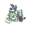
Protein / Protein/peptide , 2 types, 4 molecules ABCD
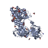
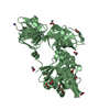
#1: Protein
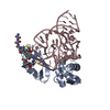
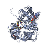
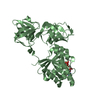
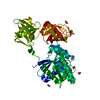
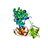
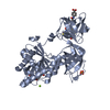
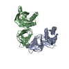
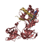
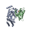
ELONGATIONFACTORTU / EF-Tu / EF-TU
Mass: 43209.270 Da / Num. of mol.: 2 / Source method: isolated from a natural source / Source: (natural) ESCHERICHIA COLI (E. coli) / References: UniProt: P0A6N1, UniProt: P0CE47*PLUS
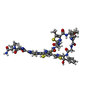
#2: Protein/peptide
THIOCILLINGE2270 / GE2270A
Type: Thiopeptide / Class: Antibiotic / Mass: 1484.681 Da / Num. of mol.: 2 / Source method: isolated from a natural source Details: GEA2270A IS A THIOPEPTIDE CONSISTING OF ONE PYRIDINE, ONE OXAZOLE AND FIVE THIAZOLE RINGS. THE OBSERVED C-TERMINAL AMINO GROUP NH2(15) IS LIKELY TO BE A POST-TRANSLATIONAL DECARBOXYLATED ...Details: GEA2270A IS A THIOPEPTIDE CONSISTING OF ONE PYRIDINE, ONE OXAZOLE AND FIVE THIAZOLE RINGS. THE OBSERVED C-TERMINAL AMINO GROUP NH2(15) IS LIKELY TO BE A POST-TRANSLATIONAL DECARBOXYLATED REMNANT OF A SER C-TERMINAL RESIDUE. Source: (natural) PLANOBISPORA ROSEA (bacteria) / References: UniProt: Q7M0J8, GE2270a
Mass: 18.015 Da / Num. of mol.: 379 / Source method: isolated from a natural source / Formula: H2O
-
Details
Compound details
GEA2270A is a member of a sulphur-rich heterocyclic peptides class. All members share a macrocylic ...GEA2270A is a member of a sulphur-rich heterocyclic peptides class. All members share a macrocylic core, consisting of a nitrogen containing, six-membered ring central to dehydroamino acids and a subset of five member ring structures including thiazoles, thiazolines and oxazoles. Post translational maturation of thiazole and oxazole containing antibiotics involves the enzymic condensation of a Cys or Ser with the alpha- carbonyl of the preceding amino acid to form a thioether or ether bond, then dehydration to form a double bond with the alpha-amino nitrogen. Thiazoline or oxazoline ring are dehydrogenated to form thiazole or oxazole rings. In addition cross-linking of a Ser and a Cys-Ser pair usually separated by 7 or 8 residues along the peptide chain produce a pyridinyl ring. The Ser residues are dehydrated to didehydroalanines, then bonded between their beta carbons. The alpha carbonyl of the Cys condenses with alpha carbon of the first Ser to form a pyridinyl ring. The ring may be mutiply dehydrogenated to form a pyridine ring with loss of the amino nitrogen of the first Ser. Several isomers are produced. The structural differences between them lie in the extent of the modifications, methylation, methoxylation, and oxidation of the thiazole and oxazole rings, and methylation of asparagine. The amidation of Pro-14 probably does not occur by the same mechanism, oxidative cleavage of glycine, as in eukaryotes.
-
Experimental details
-
Experiment
Experiment
Method: X-RAY DIFFRACTION / Number of used crystals: 1
-
Sample preparation
Crystal
Density Matthews: 2.5 Å3/Da / Density % sol: 50.84 %
In the structure databanks used in Yorodumi, some data are registered as the other names, "COVID-19 virus" and "2019-nCoV". Here are the details of the virus and the list of structure data.
Jan 31, 2019. EMDB accession codes are about to change! (news from PDBe EMDB page)
EMDB accession codes are about to change! (news from PDBe EMDB page)
The allocation of 4 digits for EMDB accession codes will soon come to an end. Whilst these codes will remain in use, new EMDB accession codes will include an additional digit and will expand incrementally as the available range of codes is exhausted. The current 4-digit format prefixed with “EMD-” (i.e. EMD-XXXX) will advance to a 5-digit format (i.e. EMD-XXXXX), and so on. It is currently estimated that the 4-digit codes will be depleted around Spring 2019, at which point the 5-digit format will come into force.
The EM Navigator/Yorodumi systems omit the EMD- prefix.
Related info.:Q: What is EMD? / ID/Accession-code notation in Yorodumi/EM Navigator
Yorodumi is a browser for structure data from EMDB, PDB, SASBDB, etc.
This page is also the successor to EM Navigator detail page, and also detail information page/front-end page for Omokage search.
The word "yorodu" (or yorozu) is an old Japanese word meaning "ten thousand". "mi" (miru) is to see.
Related info.:EMDB / PDB / SASBDB / Comparison of 3 databanks / Yorodumi Search / Aug 31, 2016. New EM Navigator & Yorodumi / Yorodumi Papers / Jmol/JSmol / Function and homology information / Changes in new EM Navigator and Yorodumi
 Movie
Movie Controller

 Yorodumi
 Open data
 Basic information
 Components
 Keywords
 THIOPEPTIDE /
  Function and homology information
 Authors
 Citation
 Structure visualization
 Downloads & links
 Other downloads
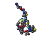

 PDBj
PDBj


 Assembly
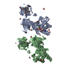
 Type: Thiopeptide


 Mass: 24.305 Da / Num. of mol.: 2 / Source method: obtained synthetically / Formula: Mg
 Type: RNA linking / Mass: 443.201 Da / Num. of mol.: 2 / Source method: obtained synthetically / Formula: C10H15N5O11P2 / Comment: GDP, energy-carrying molecule*YM
 Mass: 59.044 Da / Num. of mol.: 30 / Source method: obtained synthetically / Formula: C2H3O2
 Sample preparation
 / Beamline: BL7-1 / Wavelength: 1.08
  Processing