Mass: 18.015 Da / Num. of mol.: 244 / Source method: isolated from a natural source / Formula: H2O
-
Experimental details
-
Experiment
Experiment
Method: X-RAY DIFFRACTION / Number of used crystals: 6
-
Sample preparation
Crystal
Density Matthews: 3 Å3/Da / Density % sol: 58.4 %
Crystal grow
Method: vapor diffusion, sitting drop Details: Multiple crystals were used to collect the native and each derivative data set. VAPOR DIFFUSION, SITTING DROP
Monochromator: graphite / Protocol: SINGLE WAVELENGTH / Monochromatic (M) / Laue (L): M / Scattering type: x-ray
Radiation wavelength
Wavelength: 1.5418 Å / Relative weight: 1
Reflection
Resolution: 2.7→47.3 Å / Num. obs: 73085 / % possible obs: 92.3 % / Rmerge(I) obs: 0.099
-
Processing
Software
Name
Version
Classification
NB
CNS
refinement
PDB_EXTRACT
2
dataextraction
MERLOT
phasing
Refinement
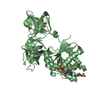
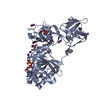
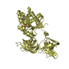
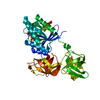
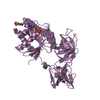
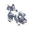
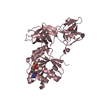
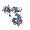
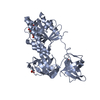
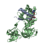
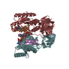
Method to determine structure: MOLECULAR REPLACEMENT Starting model: an unrefined 2.7 Angstrom model of E. coli trypsin-modified EF-Tu-MgGDP, consisting of residues 9 to 40, 50 to 258 and 260 to 393, plus one Mg ion and GDP Resolution: 2.8→40 Å / Cross valid method: THROUGHOUT / σ(F): 0 / Stereochemistry target values: Engh & Huber Details: Each molecular replacement solution was verified by successfully cross-phasing the mercury derivative sites with the generated model phases.
Rfactor
Num. reflection
% reflection
Selection details
Rfree
0.223
4601
6.5 %
random
Rwork
0.18
-
-
-
obs
-
64651
91.6 %
-
Solvent computation
Bsol: 40.313 Å2
Displacement parameters
Biso mean: 44.966 Å2
Baniso -1
Baniso -2
Baniso -3
1-
6.585 Å2
0 Å2
-7.172 Å2
2-
-
-1.468 Å2
0 Å2
3-
-
-
-5.117 Å2
Refinement step
Cycle: LAST / Resolution: 2.8→40 Å
Protein
Nucleic acid
Ligand
Solvent
Total
Num. atoms
16998
0
366
244
17608
Refine LS restraints
Refine-ID
Type
Dev ideal
Dev ideal target
X-RAY DIFFRACTION
c_mcbond_it
2.781
1.5
X-RAY DIFFRACTION
c_mcangle_it
4.366
2
X-RAY DIFFRACTION
c_bond_d
0.02
X-RAY DIFFRACTION
c_angle_deg
1.82
Xplor file
Refine-ID
Serial no
Param file
X-RAY DIFFRACTION
1
CNS_TOPPAR:protein_rep.param
X-RAY DIFFRACTION
2
MYTOPPAR:gdp96_cns.param
X-RAY DIFFRACTION
3
CNS_TOPPAR:ion.param
X-RAY DIFFRACTION
4
MYTOPPAR:tac_cns5.param
X-RAY DIFFRACTION
5
CNS_TOPPAR:water_rep.param
+
About Yorodumi
-
News
-
Feb 9, 2022. New format data for meta-information of EMDB entries
New format data for meta-information of EMDB entries
Version 3 of the EMDB header file is now the official format.
The previous official version 1.9 will be removed from the archive.
In the structure databanks used in Yorodumi, some data are registered as the other names, "COVID-19 virus" and "2019-nCoV". Here are the details of the virus and the list of structure data.
Jan 31, 2019. EMDB accession codes are about to change! (news from PDBe EMDB page)
EMDB accession codes are about to change! (news from PDBe EMDB page)
The allocation of 4 digits for EMDB accession codes will soon come to an end. Whilst these codes will remain in use, new EMDB accession codes will include an additional digit and will expand incrementally as the available range of codes is exhausted. The current 4-digit format prefixed with “EMD-” (i.e. EMD-XXXX) will advance to a 5-digit format (i.e. EMD-XXXXX), and so on. It is currently estimated that the 4-digit codes will be depleted around Spring 2019, at which point the 5-digit format will come into force.
The EM Navigator/Yorodumi systems omit the EMD- prefix.
Related info.:Q: What is EMD? / ID/Accession-code notation in Yorodumi/EM Navigator
Yorodumi is a browser for structure data from EMDB, PDB, SASBDB, etc.
This page is also the successor to EM Navigator detail page, and also detail information page/front-end page for Omokage search.
The word "yorodu" (or yorozu) is an old Japanese word meaning "ten thousand". "mi" (miru) is to see.
Related info.:EMDB / PDB / SASBDB / Comparison of 3 databanks / Yorodumi Search / Aug 31, 2016. New EM Navigator & Yorodumi / Yorodumi Papers / Jmol/JSmol / Function and homology information / Changes in new EM Navigator and Yorodumi
 Movie
Movie Controller

 Yorodumi
 Open data
 Basic information
 Components
 Keywords
 Function and homology information
 X-RAY DIFFRACTION /
  Authors
 Citation
 Structure visualization
 Downloads & links
 Other downloads
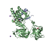

 PDBj
PDBj


 Assembly
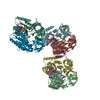


 Mass: 24.305 Da / Num. of mol.: 6 / Source method: obtained synthetically / Formula: Mg
 Type: RNA linking / Mass: 443.201 Da / Num. of mol.: 6 / Source method: obtained synthetically / Formula: C10H15N5O11P2 / Comment: GDP, energy-carrying molecule*YM
 Mass: 444.435 Da / Num. of mol.: 6 / Source method: obtained synthetically / Formula: C22H24N2O8 / Comment: antibiotic*YM
 Sample preparation
 Processing