 Movie
Movie Controller

[English] 日本語
 Yorodumi
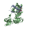
- PDB-6i8r: Crystal structure of an ancient sequence-reconstructed Elongation... -
+ Open data

- Basic information
| Entry | Database: PDB / ID: 6i8r | |||||||||||||||
|---|---|---|---|---|---|---|---|---|---|---|---|---|---|---|---|---|
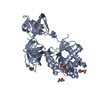
| Title | Crystal structure of an ancient sequence-reconstructed Elongation factor Tu (node 170) | |||||||||||||||
 Components Components | Elongation Factor Tu | |||||||||||||||
 Keywords Keywords | TRANSLATION / Protein Synthesis / Elongation Factor / Sequence reconstruction / ancient protein | |||||||||||||||
| Function / homology | Translation factors / Elongation Factor Tu (Ef-tu); domain 3 / P-loop containing nucleotide triphosphate hydrolases / Beta Barrel / Rossmann fold / 3-Layer(aba) Sandwich / Mainly Beta / Alpha Beta / GUANOSINE-5'-DIPHOSPHATE Function and homology information Function and homology information | |||||||||||||||
| Biological species | synthetic construct (others) | |||||||||||||||
| Method |  X-RAY DIFFRACTION / SYNCHROTRON / MOLECULAR REPLACEMENT / Resolution: 2 Å X-RAY DIFFRACTION / SYNCHROTRON / MOLECULAR REPLACEMENT / Resolution: 2 Å | |||||||||||||||
 Authors Authors | Majumdar, S. / Bergfors, T. / Sanyal, S. | |||||||||||||||
| Funding support |  Sweden, 4items Sweden, 4items
| |||||||||||||||
 Citation Citation | Journal: To be published Title: Crystal structure of an ancient sequence-reconstructed Elongation factor Tu (node 170) Authors: Majumdar, S. / Bergfors, T. / Sanyal, S. | |||||||||||||||
| History |
|
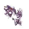
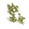
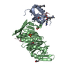
- Structure visualization
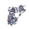
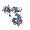
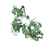
| Structure viewer | Molecule: MolmilJmol/JSmol |
|---|
- Downloads & links
-Download
| PDBx/mmCIF format | 6i8r.cif.gz | 93.2 KB | Display | PDBx/mmCIF format |
|---|---|---|---|---|
| PDB format | pdb6i8r.ent.gz | 68 KB | Display | PDB format |
| PDBx/mmJSON format | 6i8r.json.gz | Tree view | PDBx/mmJSON format | |
| Others |  Other downloads Other downloads |
-Validation report
| Arichive directory | https://data.pdbj.org/pub/pdb/validation_reports/i8/6i8rftp://data.pdbj.org/pub/pdb/validation_reports/i8/6i8r | HTTPS FTP |
|---|
-Related structure data
| Related structure data |  1dg1S S: Starting model for refinement |
|---|---|
| Similar structure data |

-Links
 PDBj
PDBj
- Assembly
| Deposited unit | 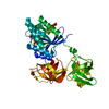
| ||||||||
|---|---|---|---|---|---|---|---|---|---|
| 1 |
| ||||||||
| Unit cell |
|
-Components
| #1: Protein | Mass: 43870.992 Da / Num. of mol.: 1 Source method: isolated from a genetically manipulated source Source: (gene. exp.) synthetic construct (others) / Production host:  |
|---|---|
| #2: Chemical | ChemComp-GDP /   Type: RNA linking / Mass: 443.201 Da / Num. of mol.: 1 / Source method: obtained synthetically / Formula: C10H15N5O11P2 / Comment: GDP, energy-carrying molecule*YM Type: RNA linking / Mass: 443.201 Da / Num. of mol.: 1 / Source method: obtained synthetically / Formula: C10H15N5O11P2 / Comment: GDP, energy-carrying molecule*YM |
| #3: Chemical | ChemComp-MG /   Mass: 24.305 Da / Num. of mol.: 1 / Source method: obtained synthetically / Formula: Mg Mass: 24.305 Da / Num. of mol.: 1 / Source method: obtained synthetically / Formula: Mg |
| #4: Water | ChemComp-HOH /  Mass: 18.015 Da / Num. of mol.: 242 / Source method: isolated from a natural source / Formula: H2O Mass: 18.015 Da / Num. of mol.: 242 / Source method: isolated from a natural source / Formula: H2O |
-Experimental details
-Experiment
| Experiment | Method: X-RAY DIFFRACTION / Number of used crystals: 1 |
|---|
- Sample preparation
| Crystal | Density Matthews: 2.99 Å3/Da / Density % sol: 58.85 % |
|---|---|
| Crystal grow | Temperature: 296 K / Method: vapor diffusion, sitting drop Details: 0.2 M Potassium Chloride, 20% w/v Polyethylene glycol 3350 |
-Data collection
| Diffraction | Mean temperature: 100 K / Serial crystal experiment: N | ||||||||||||||||||||||||
|---|---|---|---|---|---|---|---|---|---|---|---|---|---|---|---|---|---|---|---|---|---|---|---|---|---|
| Diffraction source | Source: SYNCHROTRON / Site: ESRF  / Beamline: ID23-1 / Wavelength: 0.97242 Å / Beamline: ID23-1 / Wavelength: 0.97242 Å | ||||||||||||||||||||||||
| Detector | Type: DECTRIS PILATUS 6M / Detector: PIXEL / Date: Sep 27, 2018 | ||||||||||||||||||||||||
| Radiation | Protocol: SINGLE WAVELENGTH / Monochromatic (M) / Laue (L): M / Scattering type: x-ray | ||||||||||||||||||||||||
| Radiation wavelength | Wavelength: 0.97242 Å / Relative weight: 1 | ||||||||||||||||||||||||
| Reflection | Resolution: 2→45.42 Å / Num. obs: 33564 / % possible obs: 99 % / Redundancy: 9.7 % / CC1/2: 0.999 / Rmerge(I) obs: 0.052 / Rpim(I) all: 0.018 / Rrim(I) all: 0.055 / Net I/σ(I): 17.5 / Num. measured all: 325593 | ||||||||||||||||||||||||
| Reflection shell | Diffraction-ID: 1
|
- Processing
| Software |
| ||||||||||||||||||||||||||||||||||||||||||||||||||||||||||||
|---|---|---|---|---|---|---|---|---|---|---|---|---|---|---|---|---|---|---|---|---|---|---|---|---|---|---|---|---|---|---|---|---|---|---|---|---|---|---|---|---|---|---|---|---|---|---|---|---|---|---|---|---|---|---|---|---|---|---|---|---|---|
| Refinement | Method to determine structure: MOLECULAR REPLACEMENT Starting model: 1DG1 Resolution: 2→45.42 Å / Cor.coef. Fo:Fc: 0.976 / Cor.coef. Fo:Fc free: 0.961 / SU B: 8.351 / SU ML: 0.195 / Cross valid method: THROUGHOUT / σ(F): 0 / ESU R: 0.156 / ESU R Free: 0.153 Details: HYDROGENS HAVE BEEN ADDED IN THE RIDING POSITIONS U VALUES : REFINED INDIVIDUALLY
| ||||||||||||||||||||||||||||||||||||||||||||||||||||||||||||
| Solvent computation | Ion probe radii: 0.8 Å / Shrinkage radii: 0.8 Å / VDW probe radii: 1.2 Å | ||||||||||||||||||||||||||||||||||||||||||||||||||||||||||||
| Displacement parameters | Biso max: 263.76 Å2 / Biso mean: 66.933 Å2 / Biso min: 43 Å2
| ||||||||||||||||||||||||||||||||||||||||||||||||||||||||||||
| Refinement step | Cycle: final / Resolution: 2→45.42 Å
| ||||||||||||||||||||||||||||||||||||||||||||||||||||||||||||
| Refine LS restraints |
| ||||||||||||||||||||||||||||||||||||||||||||||||||||||||||||
| LS refinement shell | Resolution: 1.997→2.049 Å / Rfactor Rfree error: 0 / Total num. of bins used: 20
|
