 Movie
Movie Controller


+ Open data

- Basic information
| Entry | Database: PDB / ID: 1bhh | ||||||
|---|---|---|---|---|---|---|---|
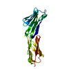
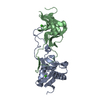
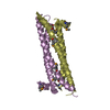
| Title | FREE P56LCK SH2 DOMAIN | ||||||
 Components Components |
| ||||||
 Keywords Keywords |  SH2 DOMAIN / PHOSPHORYLATION / TRANSFERASE SH2 DOMAIN / PHOSPHORYLATION / TRANSFERASE | ||||||
| Function / homology |  Function and homology informationregulation of lymphocyte activation / positive regulation of leukocyte cell-cell adhesion / Fc-gamma receptor signaling pathway / CD28 co-stimulation / intracellular zinc ion homeostasis / FLT3 signaling through SRC family kinases / CD4 receptor binding / Nef Mediated CD4 Down-regulation / Nef and signal transduction / Interleukin-2 signaling ...regulation of lymphocyte activation / positive regulation of leukocyte cell-cell adhesion / Fc-gamma receptor signaling pathway / CD28 co-stimulation / intracellular zinc ion homeostasis / FLT3 signaling through SRC family kinases / CD4 receptor binding / Nef Mediated CD4 Down-regulation / Nef and signal transduction / Interleukin-2 signaling / CD28 dependent Vav1 pathway / positive regulation of heterotypic cell-cell adhesion / protein serine/threonine phosphatase activity / Regulation of KIT signaling / CTLA4 inhibitory signaling / leukocyte migration / phospholipase activator activity / positive regulation of T cell receptor signaling pathway / pericentriolar material / Translocation of ZAP-70 to Immunological synapse / Phosphorylation of CD3 and TCR zeta chains / PECAM1 interactions / phospholipase binding / CD28 dependent PI3K/Akt signaling / RHOH GTPase cycle / hemopoiesis / Generation of second messenger molecules / immunological synapse / T cell differentiation / CD8 receptor binding / PD-1 signaling / phosphatidylinositol 3-kinase binding / positive regulation of intrinsic apoptotic signaling pathway / peptidyl-tyrosine autophosphorylation / release of sequestered calcium ion into cytosol / GPVI-mediated activation cascade / T cell costimulation / extrinsic component of cytoplasmic side of plasma membrane / phosphotyrosine residue binding / SH2 domain binding / T cell receptor binding / Signaling by phosphorylated juxtamembrane, extracellular and kinase domain KIT mutants / B cell receptor signaling pathway / non-specific protein-tyrosine kinase / non-membrane spanning protein tyrosine kinase activity / Signaling by SCF-KIT / platelet activation / peptidyl-tyrosine phosphorylation / cell surface receptor protein tyrosine kinase signaling pathway / activation of cysteine-type endopeptidase activity involved in apoptotic process / Constitutive Signaling by Aberrant PI3K in Cancer / positive regulation of T cell activation / Downstream TCR signaling / PIP3 activates AKT signaling / DAP12 signaling / ATPase binding / T cell receptor signaling pathway / PI5P, PP2A and IER3 Regulate PI3K/AKT Signaling / protein phosphatase binding / protein tyrosine kinase activity / intracellular signal transduction / response to xenobiotic stimulus / membrane raft / protein phosphorylation / signaling receptor binding / innate immune response / protein kinase binding / extracellular exosome / ATP binding / identical protein binding / plasma membrane / cytosol Function and homology informationregulation of lymphocyte activation / positive regulation of leukocyte cell-cell adhesion / Fc-gamma receptor signaling pathway / CD28 co-stimulation / intracellular zinc ion homeostasis / FLT3 signaling through SRC family kinases / CD4 receptor binding / Nef Mediated CD4 Down-regulation / Nef and signal transduction / Interleukin-2 signaling ...regulation of lymphocyte activation / positive regulation of leukocyte cell-cell adhesion / Fc-gamma receptor signaling pathway / CD28 co-stimulation / intracellular zinc ion homeostasis / FLT3 signaling through SRC family kinases / CD4 receptor binding / Nef Mediated CD4 Down-regulation / Nef and signal transduction / Interleukin-2 signaling / CD28 dependent Vav1 pathway / positive regulation of heterotypic cell-cell adhesion / protein serine/threonine phosphatase activity / Regulation of KIT signaling / CTLA4 inhibitory signaling / leukocyte migration / phospholipase activator activity / positive regulation of T cell receptor signaling pathway / pericentriolar material / Translocation of ZAP-70 to Immunological synapse / Phosphorylation of CD3 and TCR zeta chains / PECAM1 interactions / phospholipase binding / CD28 dependent PI3K/Akt signaling / RHOH GTPase cycle / hemopoiesis / Generation of second messenger molecules / immunological synapse / T cell differentiation / CD8 receptor binding / PD-1 signaling / phosphatidylinositol 3-kinase binding / positive regulation of intrinsic apoptotic signaling pathway / peptidyl-tyrosine autophosphorylation / release of sequestered calcium ion into cytosol / GPVI-mediated activation cascade / T cell costimulation / extrinsic component of cytoplasmic side of plasma membrane / phosphotyrosine residue binding / SH2 domain binding / T cell receptor binding / Signaling by phosphorylated juxtamembrane, extracellular and kinase domain KIT mutants / B cell receptor signaling pathway / non-specific protein-tyrosine kinase / non-membrane spanning protein tyrosine kinase activity / Signaling by SCF-KIT / platelet activation / peptidyl-tyrosine phosphorylation / cell surface receptor protein tyrosine kinase signaling pathway / activation of cysteine-type endopeptidase activity involved in apoptotic process / Constitutive Signaling by Aberrant PI3K in Cancer / positive regulation of T cell activation / Downstream TCR signaling / PIP3 activates AKT signaling / DAP12 signaling / ATPase binding / T cell receptor signaling pathway / PI5P, PP2A and IER3 Regulate PI3K/AKT Signaling / protein phosphatase binding / protein tyrosine kinase activity / intracellular signal transduction / response to xenobiotic stimulus / membrane raft / protein phosphorylation / signaling receptor binding / innate immune response / protein kinase binding / extracellular exosome / ATP binding / identical protein binding / plasma membrane / cytosolSimilarity search - Function | ||||||
| Biological species |  Homo sapiens (human) Homo sapiens (human) | ||||||
| Method | X-RAY DIFFRACTION / Resolution: 1.9 Å | ||||||
 Authors Authors | Tong, L. / Warren, T.C. / Lukas, S. / Schembri-King, J. / Betageri, R. / Proudfoot, J.R. / Jakes, S. | ||||||
 Citation Citation | Journal: J.Biol.Chem. / Year: 1998 Title: Carboxymethyl-phenylalanine as a replacement for phosphotyrosine in SH2 domain binding. Authors: Tong, L. / Warren, T.C. / Lukas, S. / Schembri-King, J. / Betageri, R. / Proudfoot, J.R. / Jakes, S. | ||||||
| History |
|
- Structure visualization
| Structure viewer | Molecule: MolmilJmol/JSmol |
|---|
- Downloads & links
-Download
| PDBx/mmCIF format | 1bhh.cif.gz | 55.1 KB | Display | PDBx/mmCIF format |
|---|---|---|---|---|
| PDB format | pdb1bhh.ent.gz | 40.2 KB | Display | PDB format |
| PDBx/mmJSON format | 1bhh.json.gz | Tree view | PDBx/mmJSON format | |
| Others |  Other downloads Other downloads |
-Validation report
| Arichive directory | https://data.pdbj.org/pub/pdb/validation_reports/bh/1bhhftp://data.pdbj.org/pub/pdb/validation_reports/bh/1bhh | HTTPS FTP |
|---|
-Related structure data
-Links
 PDBj
PDBj


- Assembly
| Deposited unit | 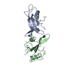
| ||||||||
|---|---|---|---|---|---|---|---|---|---|
| 1 |
| ||||||||
| Unit cell |
|
-Components
| #1: Protein | Mass: 12257.631 Da / Num. of mol.: 1 / Fragment: SH2 DOMAIN Source method: isolated from a genetically manipulated source Source: (gene. exp.) Homo sapiens (human) / Production host:  Escherichia coli (E. coli) / References: UniProt: P06239, EC: 2.7.1.112 Escherichia coli (E. coli) / References: UniProt: P06239, EC: 2.7.1.112 |
|---|---|
| #2: Protein | Mass: 11743.100 Da / Num. of mol.: 1 Source method: isolated from a genetically manipulated source Source: (gene. exp.) Homo sapiens (human) / Production host: Escherichia coli (E. coli) / References: UniProt: P06239 |
| #3: Water | ChemComp-HOH / Water Mass: 18.015 Da / Num. of mol.: 141 / Source method: isolated from a natural source / Formula: H2O Mass: 18.015 Da / Num. of mol.: 141 / Source method: isolated from a natural source / Formula: H2O |
-Experimental details
-Experiment
| Experiment | Method: X-RAY DIFFRACTION / Number of used crystals: 1 |
|---|
- Sample preparation
| Crystal | Density Matthews: 1.99 Å3/Da / Density % sol: 38.15 % | ||||||||||||||||||||||||||||||||||||||||||
|---|---|---|---|---|---|---|---|---|---|---|---|---|---|---|---|---|---|---|---|---|---|---|---|---|---|---|---|---|---|---|---|---|---|---|---|---|---|---|---|---|---|---|---|
| Crystal | *PLUS | ||||||||||||||||||||||||||||||||||||||||||
| Crystal grow | *PLUS Method: vapor diffusion, hanging drop / pH: 4.5 | ||||||||||||||||||||||||||||||||||||||||||
| Components of the solutions | *PLUS
|
-Data collection
| Diffraction | Mean temperature: 100 K |
|---|---|
| Diffraction source | Source: ROTATING ANODE / Type: RIGAKU RUH2R / Wavelength: 1.5418 |
| Detector | Type: RIGAKU / Detector: IMAGE PLATE / Date: Dec 10, 1995 |
| Radiation | Monochromator: GRAPHITE(002) / Monochromatic (M) / Laue (L): M / Scattering type: x-ray |
| Radiation wavelength | Wavelength: 1.5418 Å / Relative weight: 1 |
| Reflection | Resolution: 1.9→20 Å / Num. obs: 13477 / Observed criterion σ(I): 1 / Rmerge(I) obs: 0.06 |
| Reflection | *PLUS Num. measured all: 42626 |
- Processing
| Software |
| ||||||||||||||||||||||||||||||||||||||||||||||||||||||||||||
|---|---|---|---|---|---|---|---|---|---|---|---|---|---|---|---|---|---|---|---|---|---|---|---|---|---|---|---|---|---|---|---|---|---|---|---|---|---|---|---|---|---|---|---|---|---|---|---|---|---|---|---|---|---|---|---|---|---|---|---|---|---|
| Refinement | Resolution: 1.9→6 Å / σ(F): 2
| ||||||||||||||||||||||||||||||||||||||||||||||||||||||||||||
| Refinement step | Cycle: LAST / Resolution: 1.9→6 Å
| ||||||||||||||||||||||||||||||||||||||||||||||||||||||||||||
| Refine LS restraints |
| ||||||||||||||||||||||||||||||||||||||||||||||||||||||||||||
| Software | *PLUS Name: X-PLOR / Version: 3.1 / Classification: refinement | ||||||||||||||||||||||||||||||||||||||||||||||||||||||||||||
| Refinement | *PLUS Rfactor obs: 0.24 / Rfactor Rwork: 0.24 | ||||||||||||||||||||||||||||||||||||||||||||||||||||||||||||
| Solvent computation | *PLUS | ||||||||||||||||||||||||||||||||||||||||||||||||||||||||||||
| Displacement parameters | *PLUS |
