 Movie
Movie Controller

[English] 日本語
 Yorodumi
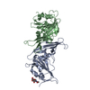
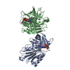
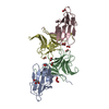
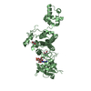
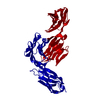
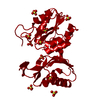
- PDB-1cdh: STRUCTURES OF AN HIV AND MHC BINDING FRAGMENT FROM HUMAN CD4 AS R... -
+ Open data

- Basic information
| Entry | Database: PDB / ID: 1cdh | |||||||||
|---|---|---|---|---|---|---|---|---|---|---|
| Title | STRUCTURES OF AN HIV AND MHC BINDING FRAGMENT FROM HUMAN CD4 AS REFINED IN TWO CRYSTAL LATTICES | |||||||||
 Components Components | T CELL SURFACE GLYCOPROTEIN CD4 | |||||||||
 Keywords Keywords | T-CELL SURFACE GLYCOPROTEIN | |||||||||
| Function / homology |  Function and homology information Function and homology informationhelper T cell enhancement of adaptive immune response / interleukin-16 binding / interleukin-16 receptor activity / maintenance of protein location in cell / cellular response to ionomycin / response to methamphetamine hydrochloride / T cell selection / MHC class II protein binding / interleukin-15-mediated signaling pathway / cellular response to granulocyte macrophage colony-stimulating factor stimulus ...helper T cell enhancement of adaptive immune response / interleukin-16 binding / interleukin-16 receptor activity / maintenance of protein location in cell / cellular response to ionomycin / response to methamphetamine hydrochloride / T cell selection / MHC class II protein binding / interleukin-15-mediated signaling pathway / cellular response to granulocyte macrophage colony-stimulating factor stimulus / positive regulation of monocyte differentiation / Alpha-defensins / Nef Mediated CD4 Down-regulation / regulation of T cell activation / response to vitamin D / Other interleukin signaling / extracellular matrix structural constituent / T cell receptor complex / enzyme-linked receptor protein signaling pathway / Translocation of ZAP-70 to Immunological synapse / Phosphorylation of CD3 and TCR zeta chains / Generation of second messenger molecules / macrophage differentiation / immunoglobulin binding / T cell differentiation / Co-inhibition by PD-1 / positive regulation of calcium ion transport into cytosol / Binding and entry of HIV virion / positive regulation of interleukin-2 production / coreceptor activity / positive regulation of T cell proliferation / positive regulation of calcium-mediated signaling / cell surface receptor protein tyrosine kinase signaling pathway / protein tyrosine kinase binding / clathrin-coated endocytic vesicle membrane / calcium-mediated signaling / Vpu mediated degradation of CD4 / MHC class II protein complex binding / response to estradiol / transmembrane signaling receptor activity / Downstream TCR signaling / Cargo recognition for clathrin-mediated endocytosis / T cell receptor signaling pathway / Clathrin-mediated endocytosis / virus receptor activity / signaling receptor activity / defense response to Gram-negative bacterium / adaptive immune response / response to ethanol / early endosome / cell surface receptor signaling pathway / cell adhesion / immune response / membrane raft / endoplasmic reticulum lumen / external side of plasma membrane / symbiont entry into host cell / lipid binding / protein kinase binding / endoplasmic reticulum membrane / enzyme binding / signal transduction / protein homodimerization activity / zinc ion binding / identical protein binding / plasma membrane Similarity search - Function | |||||||||
| Biological species |  Homo sapiens (human) Homo sapiens (human) | |||||||||
| Method |  X-RAY DIFFRACTION / Resolution: 2.3 Å X-RAY DIFFRACTION / Resolution: 2.3 Å | |||||||||
 Authors Authors | Ryu, S.E. / Truneh, A. / Sweet, R.W. / Hendrickson, W.A. | |||||||||
 Citation Citation | Journal: Structure / Year: 1994 Title: Structures of an HIV and MHC binding fragment from human CD4 as refined in two crystal lattices. Authors: Ryu, S.E. / Truneh, A. / Sweet, R.W. / Hendrickson, W.A. | |||||||||
| History |
|
- Structure visualization
| Structure viewer | Molecule: MolmilJmol/JSmol |
|---|
- Downloads & links
-Download
| PDBx/mmCIF format | 1cdh.cif.gz | 47.7 KB | Display | PDBx/mmCIF format |
|---|---|---|---|---|
| PDB format | pdb1cdh.ent.gz | 33.9 KB | Display | PDB format |
| PDBx/mmJSON format | 1cdh.json.gz | Tree view | PDBx/mmJSON format | |
| Others |  Other downloads Other downloads |
-Validation report
| Arichive directory | https://data.pdbj.org/pub/pdb/validation_reports/cd/1cdhftp://data.pdbj.org/pub/pdb/validation_reports/cd/1cdh | HTTPS FTP |
|---|
-Related structure data


-Links
 PDBj
PDBj


- Assembly
| Deposited unit | 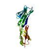
| ||||||||
|---|---|---|---|---|---|---|---|---|---|
| 1 | 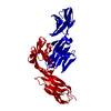
| ||||||||
| Unit cell |
|
-Components
| #1: Antibody | Mass: 19725.414 Da / Num. of mol.: 1 Source method: isolated from a genetically manipulated source Source: (gene. exp.) Homo sapiens (human) / Organ: OVARY / References: UniProt: P01730 |
|---|---|
| #2: Water | ChemComp-HOH /  Mass: 18.015 Da / Num. of mol.: 86 / Source method: isolated from a natural source / Formula: H2O Mass: 18.015 Da / Num. of mol.: 86 / Source method: isolated from a natural source / Formula: H2O |
| Has protein modification | Y |
-Experimental details
-Experiment
| Experiment | Method: X-RAY DIFFRACTION |
|---|
- Sample preparation
| Crystal | Density Matthews: 2.48 Å3/Da / Density % sol: 50.33 % | ||||||||||||||||||||
|---|---|---|---|---|---|---|---|---|---|---|---|---|---|---|---|---|---|---|---|---|---|
| Crystal grow | *PLUS Temperature: 20 ℃ / pH: 7 / Method: vapor diffusion, hanging dropDetails: referred to 'Ryu, S.-E.', (1990) Nature, 348, 419-426 | ||||||||||||||||||||
| Components of the solutions | *PLUS
|
-Data collection
| Radiation | Scattering type: x-ray |
|---|---|
| Radiation wavelength | Relative weight: 1 |
| Reflection | *PLUS Highest resolution: 2.3 Å / Lowest resolution: 10 Å / Num. all: 8479 / % possible obs: 95 % / Rmerge(I) obs: 0.086 |
- Processing
| Software |
| ||||||||||||||||||||||||||||||||||||||||||||||||||||||||||||||||||||||||||||||||
|---|---|---|---|---|---|---|---|---|---|---|---|---|---|---|---|---|---|---|---|---|---|---|---|---|---|---|---|---|---|---|---|---|---|---|---|---|---|---|---|---|---|---|---|---|---|---|---|---|---|---|---|---|---|---|---|---|---|---|---|---|---|---|---|---|---|---|---|---|---|---|---|---|---|---|---|---|---|---|---|---|---|
| Refinement | Resolution: 2.3→10 Å / σ(F): 4 /
| ||||||||||||||||||||||||||||||||||||||||||||||||||||||||||||||||||||||||||||||||
| Refinement step | Cycle: LAST / Resolution: 2.3→10 Å
| ||||||||||||||||||||||||||||||||||||||||||||||||||||||||||||||||||||||||||||||||
| Refine LS restraints |
| ||||||||||||||||||||||||||||||||||||||||||||||||||||||||||||||||||||||||||||||||
| Software | *PLUS Name: PROLSQ / Classification: refinement | ||||||||||||||||||||||||||||||||||||||||||||||||||||||||||||||||||||||||||||||||
| Refinement | *PLUS Highest resolution: 2.3 Å / Lowest resolution: 10 Å / Num. reflection obs: 7189 / σ(F): 4 / Rfactor obs: 0.193 | ||||||||||||||||||||||||||||||||||||||||||||||||||||||||||||||||||||||||||||||||
| Solvent computation | *PLUS | ||||||||||||||||||||||||||||||||||||||||||||||||||||||||||||||||||||||||||||||||
| Displacement parameters | *PLUS | ||||||||||||||||||||||||||||||||||||||||||||||||||||||||||||||||||||||||||||||||
| Refine LS restraints | *PLUS
|
