 Movie
Movie Controller

[English] 日本語
 Yorodumi
- EMDB-10896: First cryo-ET / subtomogram averaging of primary cilia microtubul... -
+ Open data

- Basic information
| Entry | Database: EMDB / ID: EMD-10896 | |||||||||
|---|---|---|---|---|---|---|---|---|---|---|
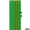
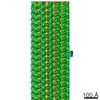
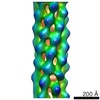
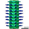
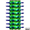
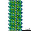
| Title | First cryo-ET / subtomogram averaging of primary cilia microtubules show that A-tubules are decorated by EB1 | |||||||||
 Map data Map data | Subtomogram average of EB1 decorated A-microtubule from MDCKII primary cilia axonemes | |||||||||
 Sample Sample |
| |||||||||
| Biological species |   Canis lupus familiaris (dog) Canis lupus familiaris (dog) | |||||||||
| Method | subtomogram averaging / cryo EM / Resolution: 18.56 Å | |||||||||
 Authors Authors | Kiesel P / Alvarez Viar G / Tsoy N / Maraspini R / Honigmann A / Pigino G | |||||||||
| Funding support | 1 items
| |||||||||
 Citation Citation | Journal: Nat Struct Mol Biol / Year: 2020 Title: The molecular structure of mammalian primary cilia revealed by cryo-electron tomography. Authors: Petra Kiesel / Gonzalo Alvarez Viar / Nikolai Tsoy / Riccardo Maraspini / Peter Gorilak / Vladimir Varga / Alf Honigmann / Gaia Pigino /    Abstract: Primary cilia are microtubule-based organelles that are important for signaling and sensing in eukaryotic cells. Unlike the thoroughly studied motile cilia, the three-dimensional architecture and ...Primary cilia are microtubule-based organelles that are important for signaling and sensing in eukaryotic cells. Unlike the thoroughly studied motile cilia, the three-dimensional architecture and molecular composition of primary cilia are largely unexplored. Yet, studying these aspects is necessary to understand how primary cilia function in health and disease. We developed an enabling method for investigating the structure of primary cilia isolated from MDCK-II cells at molecular resolution by cryo-electron tomography. We show that the textbook '9 + 0' arrangement of microtubule doublets is only present at the primary cilium base. A few microns out, the architecture changes into an unstructured bundle of EB1-decorated microtubules and actin filaments, putting an end to a long debate on the presence or absence of actin filaments in primary cilia. Our work provides a plethora of insights into the molecular structure of primary cilia and offers a methodological framework to study these important organelles. #1: Journal: Biorxiv / Year: 2020Title: The molecular structure of primary cilia revealed by cryo-electron tomography Authors: Kiesel P / Alvarez Viar G / Tsoy N / Maraspini R / Honigmann A / Pigino G | |||||||||
| History |
|
- Structure visualization
| Movie |
 Movie viewer Movie viewer |
|---|---|
| Structure viewer | EM map: SurfViewMolmilJmol/JSmol |
| Supplemental images |

- Downloads & links
-EMDB archive
| Map data | emd_10896.map.gz | 2.8 MB | EMDB map data format | |
|---|---|---|---|---|
| Header (meta data) | emd-10896-v30.xmlemd-10896.xml | 10.7 KB 10.7 KB | Display Display | EMDB header |
| Images | 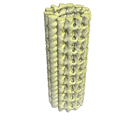 emd_10896.png emd_10896.png | 101.7 KB | ||
| Archive directory |  http://ftp.pdbj.org/pub/emdb/structures/EMD-10896ftp://ftp.pdbj.org/pub/emdb/structures/EMD-10896 http://ftp.pdbj.org/pub/emdb/structures/EMD-10896ftp://ftp.pdbj.org/pub/emdb/structures/EMD-10896 | HTTPS FTP |
-Related structure data
| Related structure data | C: citing same article ( |
|---|---|
| Similar structure data |
-Links
| EMDB pages | EMDB (EBI/PDBe) / EMDataResource |
|---|
-Map
| File | Download / File: emd_10896.map.gz / Format: CCP4 / Size: 3 MB / Type: IMAGE STORED AS FLOATING POINT NUMBER (4 BYTES) | ||||||||||||||||||||||||||||||||||||||||||||||||||||||||||||
|---|---|---|---|---|---|---|---|---|---|---|---|---|---|---|---|---|---|---|---|---|---|---|---|---|---|---|---|---|---|---|---|---|---|---|---|---|---|---|---|---|---|---|---|---|---|---|---|---|---|---|---|---|---|---|---|---|---|---|---|---|---|
| Annotation | Subtomogram average of EB1 decorated A-microtubule from MDCKII primary cilia axonemes | ||||||||||||||||||||||||||||||||||||||||||||||||||||||||||||
| Voxel size | X=Y=Z: 4.711 Å | ||||||||||||||||||||||||||||||||||||||||||||||||||||||||||||
| Density |
| ||||||||||||||||||||||||||||||||||||||||||||||||||||||||||||
| Symmetry | Space group: 1 | ||||||||||||||||||||||||||||||||||||||||||||||||||||||||||||
| Details | EMDB XML:
CCP4 map header:
| ||||||||||||||||||||||||||||||||||||||||||||||||||||||||||||
-Supplemental data
- Sample components
-Entire : Subtomogram average of EB1 decorated A-microtubule from cryo-tomo...
| Entire | Name: Subtomogram average of EB1 decorated A-microtubule from cryo-tomograms of MDCKII primary cilia |
|---|---|
| Components |
|
-Supramolecule #1: Subtomogram average of EB1 decorated A-microtubule from cryo-tomo...
| Supramolecule | Name: Subtomogram average of EB1 decorated A-microtubule from cryo-tomograms of MDCKII primary cilia type: complex / ID: 1 / Parent: 0 |
|---|---|
| Source (natural) | Organism: Canis lupus familiaris (dog) / Organ: Kidney / Tissue: tubule epithelial tissue / Organelle: primary cilium / Location in cell: axoneme |
-Experimental details
-Structure determination
| Method | cryo EM |
|---|---|
 Processing Processing | subtomogram averaging |
| Aggregation state | cell |
-Sample preparation
| Buffer | pH: 7.25 |
|---|---|
| Grid | Model: Quantifoil R3.5/1 / Material: COPPER / Mesh: 200 / Support film - Material: CARBON / Support film - topology: HOLEY / Pretreatment - Type: PLASMA CLEANING / Pretreatment - Atmosphere: AIR |
| Vitrification | Cryogen name: ETHANE / Chamber humidity: 99 % / Instrument: LEICA EM GP |
- Electron microscopy
| Microscope | FEI TITAN |
|---|---|
| Electron beam | Acceleration voltage: 300 kV / Electron source: FIELD EMISSION GUN |
| Electron optics | Illumination mode: FLOOD BEAM / Imaging mode: BRIGHT FIELDBright-field microscopy |
| Sample stage | Cooling holder cryogen: NITROGEN |
| Image recording | Film or detector model: GATAN K2 SUMMIT (4k x 4k) / Detector mode: SUPER-RESOLUTION / Average electron dose: 1.6 e/Å2 |
-Image processing
| Crystal parameters | Unit cell - A: 1 Å / Unit cell - B: 1 Å / Unit cell - C: 1 Å / Unit cell - γ: 1 ° / Unit cell - α: 1 ° / Unit cell - β: 1 ° / Space group: 1 |
|---|---|
| Extraction | Number tomograms: 10 / Number images used: 2970 |
| CTF correction | Software - Name: CTFPHASEFLIP |
| Final angle assignment | Type: OTHER |
| Final reconstruction | Resolution.type: BY AUTHOR / Resolution: 18.56 Å / Resolution method: FSC 0.143 CUT-OFF / Software - Name: PEET / Number subtomograms used: 2970 |
