 Movie
Movie Controller

[English] 日本語
 Yorodumi
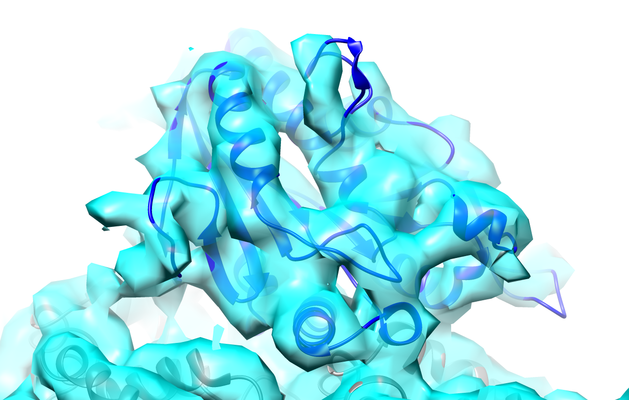
- EMDB-8408: Cryo-EM reconstruction of the yeast kinesin-5, Cin8, bound to GDP... -
+ Open data

- Basic information
| Entry | Database: EMDB / ID: EMD-8408 | |||||||||
|---|---|---|---|---|---|---|---|---|---|---|
| Title | Cryo-EM reconstruction of the yeast kinesin-5, Cin8, bound to GDP-taxol microtubules in ADP-AlFx state | |||||||||
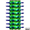
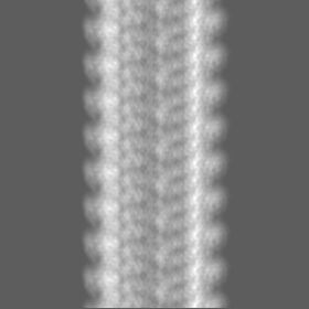
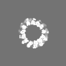
 Map data Map data | 3D helical reconstruction of Cin8 motor domain in ADP-AlFx state bound to microtubule using cryo-EM | |||||||||
 Sample Sample |
| |||||||||
| Biological species |  | |||||||||
| Method | helical reconstruction / cryo EM / Resolution: 7.3 Å | |||||||||
 Authors Authors | Cha HK | |||||||||
 Citation Citation | Journal: J Biol Chem / Year: 2017 Title: The yeast kinesin-5 Cin8 interacts with the microtubule in a noncanonical manner. Authors: Kayla M Bell / Hyo Keun Cha / Charles V Sindelar / Jared C Cochran /  Abstract: Kinesin motors play central roles in establishing and maintaining the mitotic spindle during cell division. Unlike most other kinesins, Cin8, a kinesin-5 motor in can move bidirectionally along ...Kinesin motors play central roles in establishing and maintaining the mitotic spindle during cell division. Unlike most other kinesins, Cin8, a kinesin-5 motor in can move bidirectionally along microtubules, switching directionality according to biochemical conditions, a behavior that remains largely unexplained. To this end, we used biochemical rate and equilibrium constant measurements as well as cryo-electron microscopy methodologies to investigate the microtubule interactions of the Cin8 motor domain. These experiments unexpectedly revealed that, whereas Cin8 ATPase kinetics fell within measured ranges for kinesins (especially kinesin-5 proteins), approximately four motors can bind each αβ-tubulin dimer within the microtubule lattice. This result contrasted with those observations on other known kinesins, which can bind only a single "canonical" site per tubulin dimer. Competition assays with human kinesin-5 (Eg5) only partially abrogated this behavior, indicating that Cin8 binds microtubules not only at the canonical site, but also one or more separate ("noncanonical") sites. Moreover, we found that deleting the large, class-specific insert in the microtubule-binding loop 8 reverts Cin8 to one motor per αβ-tubulin in the microtubule. The novel microtubule-binding mode of Cin8 identified here provides a potential explanation for Cin8 clustering along microtubules and potentially may contribute to the mechanism for direction reversal. | |||||||||
| History |
|
- Structure visualization
| Movie |
 Movie viewer Movie viewer |
|---|---|
| Structure viewer | EM map: SurfViewMolmilJmol/JSmol |
| Supplemental images |
- Downloads & links
-EMDB archive
| Map data | emd_8408.map.gz | 9.4 MB | EMDB map data format | |
|---|---|---|---|---|
| Header (meta data) | emd-8408-v30.xmlemd-8408.xml | 9.5 KB 9.5 KB | Display Display | EMDB header |
| FSC (resolution estimation) | emd_8408_fsc.xml | 11.6 KB | Display | FSC data file |
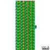
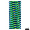
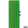
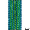
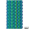
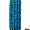
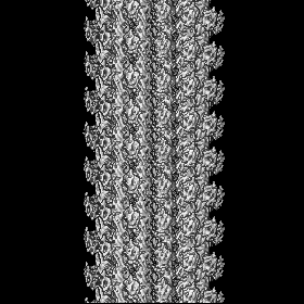
| Images |  emd_8408.png emd_8408.png | 210.4 KB | ||
| Archive directory |  http://ftp.pdbj.org/pub/emdb/structures/EMD-8408ftp://ftp.pdbj.org/pub/emdb/structures/EMD-8408 http://ftp.pdbj.org/pub/emdb/structures/EMD-8408ftp://ftp.pdbj.org/pub/emdb/structures/EMD-8408 | HTTPS FTP |
-Related structure data
| Similar structure data |
|---|
-Links
| EMDB pages | EMDB (EBI/PDBe) / EMDataResource |
|---|
-Map
| File | Download / File: emd_8408.map.gz / Format: CCP4 / Size: 83.7 MB / Type: IMAGE STORED AS FLOATING POINT NUMBER (4 BYTES) | ||||||||||||||||||||||||||||||||||||||||||||||||||||||||||||
|---|---|---|---|---|---|---|---|---|---|---|---|---|---|---|---|---|---|---|---|---|---|---|---|---|---|---|---|---|---|---|---|---|---|---|---|---|---|---|---|---|---|---|---|---|---|---|---|---|---|---|---|---|---|---|---|---|---|---|---|---|---|
| Annotation | 3D helical reconstruction of Cin8 motor domain in ADP-AlFx state bound to microtubule using cryo-EM | ||||||||||||||||||||||||||||||||||||||||||||||||||||||||||||
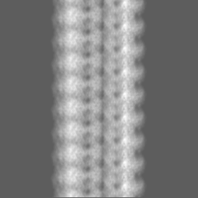
| Projections & slices | Image control
Images are generated by Spider. | ||||||||||||||||||||||||||||||||||||||||||||||||||||||||||||
| Voxel size | X=Y=Z: 2.49402 Å | ||||||||||||||||||||||||||||||||||||||||||||||||||||||||||||
| Density |
| ||||||||||||||||||||||||||||||||||||||||||||||||||||||||||||
| Symmetry | Space group: 1 | ||||||||||||||||||||||||||||||||||||||||||||||||||||||||||||
| Details | EMDB XML:
CCP4 map header:
| ||||||||||||||||||||||||||||||||||||||||||||||||||||||||||||
 Z (Sec.)
 Y (Row.)
 X (Col.)


-Supplemental data
- Sample components
-Entire : Microtubule-bound kinesin-5 Cin8 in ADP-AlFx state
| Entire | Name: Microtubule-bound kinesin-5 Cin8 in ADP-AlFx state |
|---|---|
| Components |
|
-Supramolecule #1: Microtubule-bound kinesin-5 Cin8 in ADP-AlFx state
| Supramolecule | Name: Microtubule-bound kinesin-5 Cin8 in ADP-AlFx state / type: complex / ID: 1 / Parent: 0 Details: Cin8-his monomer (residues 70-535) Taxol-stabilized microtubules |
|---|---|
| Source (natural) | Organism: |
| Recombinant expression | Organism:  |
-Experimental details
-Structure determination
| Method | cryo EM |
|---|---|
 Processing Processing | helical reconstruction |
| Aggregation state | filament |
-Sample preparation
| Buffer | pH: 6.8 Component:
Details: adjusted to pH 6.8 with KOH | |||||||||||||||
|---|---|---|---|---|---|---|---|---|---|---|---|---|---|---|---|---|
| Vitrification | Cryogen name: ETHANE / Instrument: HOMEMADE PLUNGER |
- Electron microscopy
| Microscope | FEI TECNAI F20 |
|---|---|
| Image recording | Film or detector model: GATAN K2 SUMMIT (4k x 4k) / Average electron dose: 1.71 e/Å2 |
| Electron beam | Acceleration voltage: 200 kV / Electron source:  FIELD EMISSION GUN FIELD EMISSION GUN |
| Electron optics | Illumination mode: FLOOD BEAM / Imaging mode: BRIGHT FIELD |
| Experimental equipment |  Model: Tecnai F20 / Image courtesy: FEI Company |
-Image processing
| Final reconstruction | Applied symmetry - Helical parameters - Δz: 9.455 Å Applied symmetry - Helical parameters - Δ&Phi: -25.77 ° Applied symmetry - Helical parameters - Axial symmetry: C1 (asymmetric) Resolution.type: BY AUTHOR / Resolution: 7.3 Å / Resolution method: FSC 0.143 CUT-OFF / Software - Name: FREALIGN / Number images used: 2282 |
|---|---|
| CTF correction | Software - Name: CTFFIND3 |
| Final angle assignment | Type: NOT APPLICABLE / Software - Name: SPIDER |
| FSC plot (resolution estimation) | 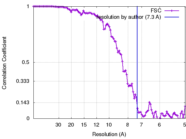 |
