 ムービー
ムービー コントローラー

+ データを開く

- 基本情報
基本情報
| 登録情報 | データベース: SASBDB / ID: SASDDE6 |
|---|---|
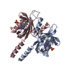
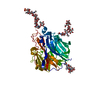
 試料 試料 | Photoreceptor sensory box 1 light-state (SB1-LOV-R66I)
|
| 機能・相同性 |  機能・相同性情報 機能・相同性情報 |
| 生物種 |  Pseudomonas putida (strain ATCC 47054 / DSM 6125 / NCIMB 11950 / KT2440) (バクテリア) Pseudomonas putida (strain ATCC 47054 / DSM 6125 / NCIMB 11950 / KT2440) (バクテリア) |
 引用 引用 | ジャーナル: PLoS One / 年: 2018 タイトル: Small-angle X-ray scattering study of the kinetics of light-dark transition in a LOV protein. 著者: Katrin Röllen / Joachim Granzin / Renu Batra-Safferling / Andreas Maximilian Stadler /  要旨: Light, oxygen, voltage (LOV) photoreceptors consist of conserved photo-responsive domains in bacteria, archaea, plants and fungi, and detect blue-light via a flavin cofactor. We investigated the blue- ...Light, oxygen, voltage (LOV) photoreceptors consist of conserved photo-responsive domains in bacteria, archaea, plants and fungi, and detect blue-light via a flavin cofactor. We investigated the blue-light induced conformational transition of the dimeric photoreceptor PpSB1-LOV-R66I from Pseudomonas putida in solution by using small-angle X-ray scattering (SAXS). SAXS experiments of the fully populated light- and dark-states under steady-state conditions revealed significant structural differences between the two states that are in agreement with the known structures determined by crystallography. We followed the transition from the light- to the dark-state by using SAXS measurements in real-time. A two-state model based on the light- and dark-state conformations could describe the measured time-course SAXS data with a relaxation time τREC of ~ 34 to 35 min being larger than the recovery time found with UV/vis spectroscopy. Unlike the flavin chromophore-based UV/vis method that is sensitive to the local chromophore environment in flavoproteins, SAXS-based assay depends on protein conformational changes and provides with an alternative to measure the recovery kinetics. |
 登録者 登録者 |
|
- 構造の表示
構造の表示
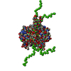
| 構造ビューア | 分子: MolmilJmol/JSmol |
|---|
- ダウンロードとリンク
 SASDDE6


-モデル
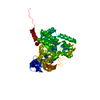
| モデル #1970 |  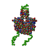 タイプ: mix / ダミー原子の半径: 1.90 A / カイ2乗値: 1.891  Omokage検索でこの集合体の類似形状データを探す (詳細) Omokage検索でこの集合体の類似形状データを探す (詳細) |
|---|---|
| モデル #1971 |   タイプ: mix / ソフトウェア: (2.8.3) / ダミー原子の半径: 1.90 A / カイ2乗値: 5.639 Omokage検索でこの集合体の類似形状データを探す (詳細) |
| モデル #1972 | 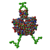 タイプ: mix / ダミー原子の半径: 1.90 A / カイ2乗値: 5.639 Omokage検索でこの集合体の類似形状データを探す (詳細) |
| モデル #1973 |   タイプ: dummy / ソフトウェア: (2.8.3) / ダミー原子の半径: 2.00 A / 対称性: P2 / カイ2乗値: 0.783 Omokage検索でこの集合体の類似形状データを探す (詳細) |
-試料
| 試料 | 名称: Photoreceptor sensory box 1 light-state (SB1-LOV-R66I) 試料濃度: 1.05-6.10 |
|---|---|
| バッファ | 名称: 10mM Tris, 10 mM NaCl / pH: 7 |
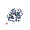
| 要素 #1060 | 名称: PpSB1-LOV-R66I light / タイプ: protein / 記述: Sensory box protein light-state (R66I) / 分子量: 18.588 / 分子数: 2 由来: Pseudomonas putida (strain ATCC 47054 / DSM 6125 / NCIMB 11950 / KT2440) 参照: UniProt: Q88E39 配列: MGSSHHHHHH SSGLVPRGSH MINAQLLQSM VDASNDGIVV AEKEGDDTIL IYVNAAFEYL TGYSRDEILY QDCRFLQGDD RDQLGRARIR KAMAEGRPCR EVLRNYRKDG SAFWNELSIT PVKSDFDQRT YFIGIQKDVS RQVELERELA ELRARPKPDE RA |
-実験情報
| ビーム | 設備名称: ESRF BM29 / 地域: Grenoble / 国: France  / 線源: X-ray synchrotron / 波長: 0.099 Å / スペクトロメータ・検出器間距離: 2.867 mm / 線源: X-ray synchrotron / 波長: 0.099 Å / スペクトロメータ・検出器間距離: 2.867 mm | ||||||||||||||||||||||||||||||||||||
|---|---|---|---|---|---|---|---|---|---|---|---|---|---|---|---|---|---|---|---|---|---|---|---|---|---|---|---|---|---|---|---|---|---|---|---|---|---|
| 検出器 | 名称: Pilatus 1M / Pixsize x: 172 mm | ||||||||||||||||||||||||||||||||||||
| スキャン |  測定日: 2014年12月2日 / 保管温度: 10 °C / セル温度: 10 °C / 照射時間: 1 sec. / フレーム数: 10 / 単位: 1/nm /
| ||||||||||||||||||||||||||||||||||||
| 距離分布関数 P(R) | 
| ||||||||||||||||||||||||||||||||||||
| 結果 | 
|
