 Movie
Movie Controller

[English] 日本語
 Yorodumi
- PDB-9xa4: Structure of the Omicron BA.4/5 Spike N-terminal domain(NTD) in c... -
+ Open data

- Basic information
| Entry | Database: PDB / ID: 9xa4 | ||||||
|---|---|---|---|---|---|---|---|
| Title | Structure of the Omicron BA.4/5 Spike N-terminal domain(NTD) in complex with the AC2 Fab | ||||||
 Components Components |
| ||||||
 Keywords Keywords | IMMUNE SYSTEM / Complex | ||||||
| Function / homology |  Function and homology information Function and homology informationsymbiont-mediated disruption of host tissue / Maturation of spike protein / host cell surface / Translation of Structural Proteins / Virion Assembly and Release / Lectin pathway of complement activation / host extracellular region / symbiont-mediated-mediated suppression of host tetherin activity / structural constituent of virion / Induction of Cell-Cell Fusion ...symbiont-mediated disruption of host tissue / Maturation of spike protein / host cell surface / Translation of Structural Proteins / Virion Assembly and Release / Lectin pathway of complement activation / host extracellular region / symbiont-mediated-mediated suppression of host tetherin activity / structural constituent of virion / Induction of Cell-Cell Fusion / positive regulation of viral entry into host cell / Initial triggering of complement / membrane fusion / host cell endoplasmic reticulum-Golgi intermediate compartment membrane / Attachment and Entry / entry receptor-mediated virion attachment to host cell / receptor-mediated virion attachment to host cell / host cell surface receptor binding / symbiont-mediated suppression of host innate immune response / endocytosis involved in viral entry into host cell / receptor ligand activity / fusion of virus membrane with host plasma membrane / fusion of virus membrane with host endosome membrane / viral envelope / symbiont entry into host cell / virion attachment to host cell / host cell plasma membrane / SARS-CoV-2 activates/modulates innate and adaptive immune responses / virion membrane / membrane / identical protein binding / plasma membrane Similarity search - Function | ||||||
| Biological species |  Homo sapiens (human) Homo sapiens (human) 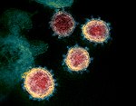 Severe acute respiratory syndrome coronavirus 2 Severe acute respiratory syndrome coronavirus 2 | ||||||
| Method |  X-RAY DIFFRACTION / SYNCHROTRON / MOLECULAR REPLACEMENT / Resolution: 2.52 Å X-RAY DIFFRACTION / SYNCHROTRON / MOLECULAR REPLACEMENT / Resolution: 2.52 Å | ||||||
 Authors Authors | Zhou, J.J. / Gao, G.F. | ||||||
| Funding support |  China, 1items China, 1items
| ||||||
 Citation Citation | Journal: Proc Natl Acad Sci U S A / Year: 2026 Title: A structural and mechanistic atlas of NTD antibody neutralization and immune escape across SARS-CoV-2 prototype and its (sub-)variants. Authors: Jianjie Zhou / Wenyu Li / Xiaoyun Wang / Junqing Sun / Shuxin Guo / Xiaoyu Rong / Zhou Tong / Lianpan Dai / William Jun Liu / Jianxun Qi / George Fu Gao / Qihui Wang / Abstract: The N-terminal domain (NTD) of the SARS-CoV-2 spike (S) is a critical antibody target, yet its epitope organization, neutralization mechanisms, and immune evasion strategies remain incompletely ...The N-terminal domain (NTD) of the SARS-CoV-2 spike (S) is a critical antibody target, yet its epitope organization, neutralization mechanisms, and immune evasion strategies remain incompletely resolved. Here, we classify NTD antibodies into nine spatially distinct classes (designated as NTD-1 to NTD-9), including a cryptic epitope defined here (NTD-8). Mechanistic studies reveal that NTD-5 and NTD-9 antibodies neutralize by inducing S1 shedding, thereby extending this mechanism to selected NTD-directed antibodies. Format profiling shows that while most NTD antibodies require bivalency, selected antibodies from NTD-3, NTD-5, and NTD-9 retain neutralizing activity in Fab form. Profiling 41 antibodies across prototype, Delta, and 17 Omicron subvariants defines an epitope-resolved escape landscape and enables dissection of three convergent evasion strategies: contact residue disruption, glycan shielding, and conformational remodeling. Notably, the KP.3.1.1 subvariant uses a dual escape mechanism in which ∆S31 introduces N30 glycosylation and substantially remodels the S27-R34 region, undermining recognition by both NTD-5 and NTD-9 antibodies. These findings provide a structural and mechanistic framework for rational vaccine and antibody design resilient to antigenic drift. | ||||||
| History |
|
- Structure visualization
| Structure viewer | Molecule: MolmilJmol/JSmol |
|---|
- Downloads & links
-Download
| PDBx/mmCIF format | 9xa4.cif.gz | 160.9 KB | Display | PDBx/mmCIF format |
|---|---|---|---|---|
| PDB format | pdb9xa4.ent.gz | 122.4 KB | Display | PDB format |
| PDBx/mmJSON format | 9xa4.json.gz | Tree view | PDBx/mmJSON format | |
| Others |  Other downloads Other downloads |
-Validation report
| Arichive directory | https://data.pdbj.org/pub/pdb/validation_reports/xa/9xa4ftp://data.pdbj.org/pub/pdb/validation_reports/xa/9xa4 | HTTPS FTP |
|---|
-Related structure data
| Related structure data |  9w6uC  9wigC  9x9zC  9xa0C  9xa5C  9xarC  9xawC C: citing same article ( |
|---|---|
| Similar structure data |


-Links
 PDBj
PDBj


- Assembly
| Deposited unit | 
| ||||||||||
|---|---|---|---|---|---|---|---|---|---|---|---|
| 1 |
| ||||||||||
| Unit cell |
|
-Components
-Protein , 1 types, 1 molecules A
| #3: Protein | Mass: 33497.645 Da / Num. of mol.: 1 / Fragment: NTD Source method: isolated from a genetically manipulated source Source: (gene. exp.) Severe acute respiratory syndrome coronavirus 2Strain: BA.4/5 / Gene: S, 2 / Production host: Homo sapiens (human) / References: UniProt: P0DTC2 |
|---|
-Antibody , 2 types, 2 molecules LH
| #1: Antibody | Mass: 22946.256 Da / Num. of mol.: 1 Source method: isolated from a genetically manipulated source Source: (gene. exp.) Homo sapiens (human) / Production host: Homo sapiens (human) |
|---|---|
| #2: Antibody | Mass: 26098.443 Da / Num. of mol.: 1 Source method: isolated from a genetically manipulated source Source: (gene. exp.) Homo sapiens (human) / Production host: Homo sapiens (human) |
-Sugars , 3 types, 3 molecules 
| #4: Polysaccharide | 2-acetamido-2-deoxy-beta-D-glucopyranose-(1-4)-2-acetamido-2-deoxy-beta-D-glucopyranose Source method: isolated from a genetically manipulated source |
|---|---|
| #5: Polysaccharide | beta-D-mannopyranose-(1-4)-2-acetamido-2-deoxy-beta-D-glucopyranose-(1-4)-2-acetamido-2-deoxy-beta- ...beta-D-mannopyranose-(1-4)-2-acetamido-2-deoxy-beta-D-glucopyranose-(1-4)-2-acetamido-2-deoxy-beta-D-glucopyranose Source method: isolated from a genetically manipulated source |
| #7: Sugar | ChemComp-NAG /  Type: D-saccharide, beta linking / Mass: 221.208 Da / Num. of mol.: 1 / Source method: obtained synthetically / Formula: C8H15NO6 Type: D-saccharide, beta linking / Mass: 221.208 Da / Num. of mol.: 1 / Source method: obtained synthetically / Formula: C8H15NO6 |
-Non-polymers , 2 types, 111 molecules 

| #6: Chemical | ChemComp-EDO / 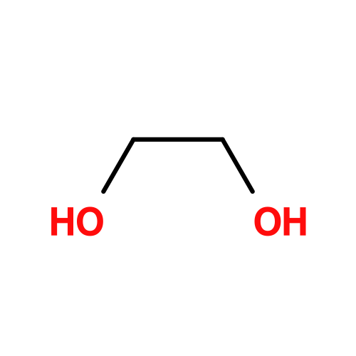 Mass: 62.068 Da / Num. of mol.: 21 / Source method: obtained synthetically / Formula: C2H6O2 Mass: 62.068 Da / Num. of mol.: 21 / Source method: obtained synthetically / Formula: C2H6O2#8: Water | ChemComp-HOH / | Mass: 18.015 Da / Num. of mol.: 90 / Source method: isolated from a natural source / Formula: H2O |
|---|
-Details
| Has ligand of interest | N |
|---|---|
| Has protein modification | Y |
-Experimental details
-Experiment
| Experiment | Method: X-RAY DIFFRACTION / Number of used crystals: 1 |
|---|
- Sample preparation
| Crystal | Density Matthews: 3.49 Å3/Da / Density % sol: 64.8 % |
|---|---|
| Crystal grow | Temperature: 289 K / Method: vapor diffusion, sitting drop Details: 0.17 M Lithium sulfate monohydrate, 0.085 M TRIS hydrochloride pH 8.5, 25.5% w/v Polyethylene glycol 4,000, 15% v/v Glycerol |
-Data collection
| Diffraction | Mean temperature: 100 K / Serial crystal experiment: N |
|---|---|
| Diffraction source | Source: SYNCHROTRON / Site: SSRF / Beamline: BL02U1 / Wavelength: 0.97925 Å |
| Detector | Type: DECTRIS EIGER2 S 9M / Detector: PIXEL / Date: Sep 15, 2024 |
| Radiation | Protocol: SINGLE WAVELENGTH / Monochromatic (M) / Laue (L): M / Scattering type: x-ray |
| Radiation wavelength | Wavelength: 0.97925 Å / Relative weight: 1 |
| Reflection | Resolution: 2.52→52.87 Å / Num. obs: 39178 / % possible obs: 99.5 % / Redundancy: 6.8 % / Biso Wilson estimate: 52.36 Å2 / CC1/2: 0.996 / Net I/σ(I): 11 |
| Reflection shell | Resolution: 2.52→2.59 Å / Num. unique obs: 2905 / CC1/2: 0.487 |
- Processing
| Software |
| |||||||||||||||||||||||||||||||||||||||||||||||||||||||||||||||||||||||||||||||||||||||||||||||||||||||||
|---|---|---|---|---|---|---|---|---|---|---|---|---|---|---|---|---|---|---|---|---|---|---|---|---|---|---|---|---|---|---|---|---|---|---|---|---|---|---|---|---|---|---|---|---|---|---|---|---|---|---|---|---|---|---|---|---|---|---|---|---|---|---|---|---|---|---|---|---|---|---|---|---|---|---|---|---|---|---|---|---|---|---|---|---|---|---|---|---|---|---|---|---|---|---|---|---|---|---|---|---|---|---|---|---|---|---|
| Refinement | Method to determine structure: MOLECULAR REPLACEMENT / Resolution: 2.52→52.87 Å / SU ML: 0.3 / Cross valid method: FREE R-VALUE / σ(F): 1.34 / Phase error: 25.54 / Stereochemistry target values: ML
| |||||||||||||||||||||||||||||||||||||||||||||||||||||||||||||||||||||||||||||||||||||||||||||||||||||||||
| Solvent computation | Shrinkage radii: 0.9 Å / VDW probe radii: 1.1 Å / Solvent model: FLAT BULK SOLVENT MODEL | |||||||||||||||||||||||||||||||||||||||||||||||||||||||||||||||||||||||||||||||||||||||||||||||||||||||||
| Refinement step | Cycle: LAST / Resolution: 2.52→52.87 Å
| |||||||||||||||||||||||||||||||||||||||||||||||||||||||||||||||||||||||||||||||||||||||||||||||||||||||||
| Refine LS restraints |
| |||||||||||||||||||||||||||||||||||||||||||||||||||||||||||||||||||||||||||||||||||||||||||||||||||||||||
| LS refinement shell |
|
