 Movie
Movie Controller


+ Open data

- Basic information
| Entry | Database: PDB / ID: 9wqk | ||||||||||||||||||
|---|---|---|---|---|---|---|---|---|---|---|---|---|---|---|---|---|---|---|---|
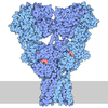
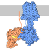
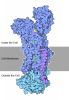
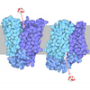
| Title | GRM5-Gi Complex Structure | ||||||||||||||||||
 Components Components |
| ||||||||||||||||||
 Keywords Keywords | MEMBRANE PROTEIN / Receptor complex | ||||||||||||||||||
| Function / homology |  Function and homology information Function and homology informationA2A adenosine receptor binding / sensory perception of hot stimulus / negative regulation of dendritic spine morphogenesis / : / G protein-coupled receptor activity involved in regulation of postsynaptic membrane potential / adenylate cyclase inhibiting G protein-coupled glutamate receptor activity / positive regulation of cellular response to hypoxia / protein localization to nuclear inner membrane / operant conditioning / phospholipase C-activating G protein-coupled glutamate receptor signaling pathway ...A2A adenosine receptor binding / sensory perception of hot stimulus / negative regulation of dendritic spine morphogenesis / : / G protein-coupled receptor activity involved in regulation of postsynaptic membrane potential / adenylate cyclase inhibiting G protein-coupled glutamate receptor activity / positive regulation of cellular response to hypoxia / protein localization to nuclear inner membrane / operant conditioning / phospholipase C-activating G protein-coupled glutamate receptor signaling pathway / positive regulation of long-term neuronal synaptic plasticity / positive regulation of sensory perception of pain / negative regulation of excitatory postsynaptic potential / positive regulation of dopamine secretion / desensitization of G protein-coupled receptor signaling pathway / G protein-coupled glutamate receptor signaling pathway / positive regulation of neural precursor cell proliferation / Class C/3 (Metabotropic glutamate/pheromone receptors) / glutamate receptor activity / nuclear inner membrane / Neurexins and neuroligins / astrocyte projection / response to morphine / response to corticosterone / temperature homeostasis / protein tyrosine kinase activator activity / conditioned place preference / regulation of synaptic transmission, glutamatergic / positive regulation of calcium-mediated signaling / regulation of eating behavior / regulation of long-term synaptic depression / adenylate cyclase inhibitor activity / T cell migration / positive regulation of protein localization to cell cortex / positive regulation of relaxation of smooth muscle / Adenylate cyclase inhibitory pathway / D2 dopamine receptor binding / response to amphetamine / adenylate cyclase-inhibiting serotonin receptor signaling pathway / G protein-coupled serotonin receptor binding / protein tyrosine kinase binding / cellular response to forskolin / locomotory behavior / regulation of mitotic spindle organization / dendritic shaft / mast cell degranulation / chemokine-mediated signaling pathway / neuropeptide signaling pathway / Regulation of insulin secretion / response to prostaglandin E / positive regulation of cholesterol biosynthetic process / synapse organization / postsynaptic density membrane / G protein-coupled receptor binding / cognition / response to peptide hormone / Schaffer collateral - CA1 synapse / cellular response to amyloid-beta / G protein-coupled receptor activity / G-protein beta/gamma-subunit complex binding / adenylate cyclase-inhibiting G protein-coupled receptor signaling pathway / adenylate cyclase-modulating G protein-coupled receptor signaling pathway / Olfactory Signaling Pathway / Activation of the phototransduction cascade / G protein-coupled acetylcholine receptor signaling pathway / G beta:gamma signalling through PLC beta / Presynaptic function of Kainate receptors / Thromboxane signalling through TP receptor / Activation of G protein gated Potassium channels / Inhibition of voltage gated Ca2+ channels via Gbeta/gamma subunits / G-protein activation / Glucagon signaling in metabolic regulation / G beta:gamma signalling through CDC42 / Prostacyclin signalling through prostacyclin receptor / Synthesis, secretion, and inactivation of Glucagon-like Peptide-1 (GLP-1) / GDP binding / G beta:gamma signalling through BTK / photoreceptor disc membrane / ADP signalling through P2Y purinoceptor 12 / Glucagon-type ligand receptors / Sensory perception of sweet, bitter, and umami (glutamate) taste / Adrenaline,noradrenaline inhibits insulin secretion / Vasopressin regulates renal water homeostasis via Aquaporins / Glucagon-like Peptide-1 (GLP1) regulates insulin secretion / G alpha (z) signalling events / cellular response to catecholamine stimulus / ADP signalling through P2Y purinoceptor 1 / G beta:gamma signalling through PI3Kgamma / ADORA2B mediated anti-inflammatory cytokines production / adenylate cyclase-activating dopamine receptor signaling pathway / cellular response to prostaglandin E stimulus / Cooperation of PDCL (PhLP1) and TRiC/CCT in G-protein beta folding / GPER1 signaling / heterotrimeric G-protein complex / Inactivation, recovery and regulation of the phototransduction cascade / G alpha (12/13) signalling events / G-protein beta-subunit binding / extracellular vesicle / positive regulation of cytosolic calcium ion concentration / Thrombin signalling through proteinase activated receptors (PARs) Similarity search - Function | ||||||||||||||||||
| Biological species |  Homo sapiens (human) Homo sapiens (human)  Mus musculus (house mouse) Mus musculus (house mouse) | ||||||||||||||||||
| Method | ELECTRON MICROSCOPY / single particle reconstruction / cryo EM / Resolution: 3.7 Å | ||||||||||||||||||
 Authors Authors | Lu, Y. / Wen, T.L. / Shen, Y.Q. / Yang, X. | ||||||||||||||||||
| Funding support |  China, 1items China, 1items
| ||||||||||||||||||
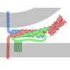
 Citation Citation | Journal: Sci Adv / Year: 2026 Title: G protein selectivity in group I metabotropic glutamate receptors. Authors: Yue Lu / Tianlei Wen / Xuhang Lu / Guimin Zhang / Tingting Meng / Tianjin Liu / Xinyan Wang / Yuequan Shen / Xue Yang / Abstract: Metabotropic glutamate (mGlu) receptors are class C G protein-coupled receptor involved in synaptic transmission and neurological disorders. Group I mGlu receptors (mGlu1 and mGlu5) predominantly ...Metabotropic glutamate (mGlu) receptors are class C G protein-coupled receptor involved in synaptic transmission and neurological disorders. Group I mGlu receptors (mGlu1 and mGlu5) predominantly couple to G, whereas group II and III receptors primarily engage G. Although G-coupling mechanisms have been defined for several group II/III receptors, how group I receptors preferentially engage G remains unclear. Here we report cryo-electron microscopy structures of active mGlu-G protein complexes (mGlu1-G, mGlu1-G, mGlu5-G, and mGlu5-G) bound to l-glutamate and positive allosteric modulators (PAMs), together with two additional activated-state structures of mGlu1. Comparative structural and biochemical analyses identify a group I-specific ICL2 insertion that promotes preferential G engagement. Each receptor dimer asymmetrically binds one G protein heterotrimer via an intracellular pocket engaging the Gα amino-terminal helix. PAM binding to one 7TM domain induces W rotation and TM6 outward movement, bringing the two 7TMs into closer. These findings provide a structural basis for preferential G engagement and activation of group I mGlu receptors. | ||||||||||||||||||
| History |
|
- Structure visualization
| Structure viewer | Molecule: MolmilJmol/JSmol |
|---|
- Downloads & links
-Download
| PDBx/mmCIF format | 9wqk.cif.gz | 445.4 KB | Display | PDBx/mmCIF format |
|---|---|---|---|---|
| PDB format | pdb9wqk.ent.gz | 353.1 KB | Display | PDB format |
| PDBx/mmJSON format | 9wqk.json.gz | Tree view | PDBx/mmJSON format | |
| Others |  Other downloads Other downloads |
-Validation report
| Arichive directory | https://data.pdbj.org/pub/pdb/validation_reports/wq/9wqkftp://data.pdbj.org/pub/pdb/validation_reports/wq/9wqk | HTTPS FTP |
|---|
-Related structure data
| Related structure data |  66174MC  27tmC  9wqlC  9wqmC  9wqnC  9wqoC M: map data used to model this data C: citing same article ( |
|---|---|
| Similar structure data |


-Links
 PDBj
PDBj






















- Assembly
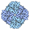
| Deposited unit | 
|
|---|---|
| 1 |
|
-Components
-Guanine nucleotide-binding protein ... , 3 types, 3 molecules ABG
| #2: Protein | Mass: 40414.047 Da / Num. of mol.: 1 Source method: isolated from a genetically manipulated source Source: (gene. exp.) Homo sapiens (human) / Gene: GNAI1 / Production host:   Spodoptera frugiperda (fall armyworm) Spodoptera frugiperda (fall armyworm)References: UniProt: P63096, Hydrolases; Acting on acid anhydrides; Acting on GTP to facilitate cellular and subcellular movement |
|---|---|
| #3: Protein | Mass: 37416.930 Da / Num. of mol.: 1 Source method: isolated from a genetically manipulated source Source: (gene. exp.) Homo sapiens (human) / Gene: GNB1 / Production host: Spodoptera frugiperda (fall armyworm) / References: UniProt: P62873 |
| #4: Protein | Mass: 8506.765 Da / Num. of mol.: 1 Source method: isolated from a genetically manipulated source Source: (gene. exp.) Homo sapiens (human) / Gene: GNG2 / Production host: Spodoptera frugiperda (fall armyworm) / References: UniProt: P59768 |
-Protein / Antibody / Sugars , 3 types, 11 molecules XYF
| #1: Protein | Mass: 95310.016 Da / Num. of mol.: 2 Source method: isolated from a genetically manipulated source Source: (gene. exp.) Homo sapiens (human) / Gene: GRM5, GPRC1E, MGLUR5 / Production host: Spodoptera frugiperda (fall armyworm) / References: UniProt: P41594#5: Antibody | | Mass: 27720.795 Da / Num. of mol.: 1 Source method: isolated from a genetically manipulated source Source: (gene. exp.) Mus musculus (house mouse) / Production host: Homo sapiens (human)#8: Sugar | ChemComp-NAG /  Type: D-saccharide, beta linking / Mass: 221.208 Da / Num. of mol.: 8 / Source method: obtained synthetically / Formula: C8H15NO6 Type: D-saccharide, beta linking / Mass: 221.208 Da / Num. of mol.: 8 / Source method: obtained synthetically / Formula: C8H15NO6 |
|---|
-Non-polymers , 4 types, 13 molecules 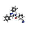

| #6: Chemical | ChemComp-YKU / 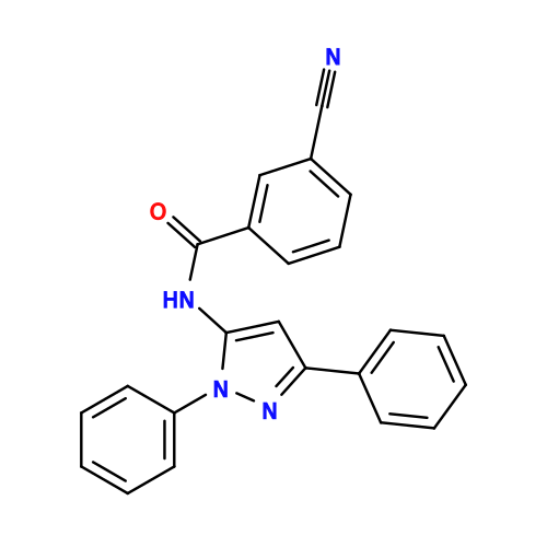 Mass: 364.399 Da / Num. of mol.: 1 / Source method: obtained synthetically / Formula: C23H16N4O / Comment: antipsychotic*YM Mass: 364.399 Da / Num. of mol.: 1 / Source method: obtained synthetically / Formula: C23H16N4O / Comment: antipsychotic*YM | ||||
|---|---|---|---|---|---|
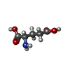
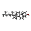
| #7: Chemical |  Type: L-gamma-peptide, C-delta linking / Mass: 147.129 Da / Num. of mol.: 2 / Source method: obtained synthetically / Formula: C5H9NO4 Type: L-gamma-peptide, C-delta linking / Mass: 147.129 Da / Num. of mol.: 2 / Source method: obtained synthetically / Formula: C5H9NO4#9: Chemical |  Mass: 35.453 Da / Num. of mol.: 2 / Source method: obtained synthetically / Formula: Cl Mass: 35.453 Da / Num. of mol.: 2 / Source method: obtained synthetically / Formula: Cl#10: Chemical | ChemComp-CLR / 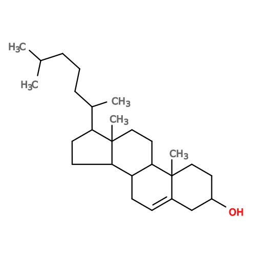 Mass: 386.654 Da / Num. of mol.: 8 / Source method: obtained synthetically / Formula: C27H46O Mass: 386.654 Da / Num. of mol.: 8 / Source method: obtained synthetically / Formula: C27H46O |
-Details
| Has ligand of interest | N |
|---|---|
| Has protein modification | Y |
-Experimental details
-Experiment
| Experiment | Method: ELECTRON MICROSCOPY |
|---|---|
| EM experiment | Aggregation state: PARTICLE / 3D reconstruction method: single particle reconstruction |
- Sample preparation
| Component | Name: GRM5-Gi complex / Type: COMPLEX / Entity ID: #1-#5 / Source: RECOMBINANT |
|---|---|
| Molecular weight | Value: 0.3 MDa / Experimental value: YES |
| Source (natural) | Organism: Homo sapiens (human) |
| Source (recombinant) | Organism: Spodoptera frugiperda (fall armyworm) |
| Buffer solution | pH: 7.2 |
| Specimen | Conc.: 10 mg/ml / Embedding applied: NO / Shadowing applied: NO / Staining applied: NO / Vitrification applied: YES |
| Vitrification | Cryogen name: ETHANE / Humidity: 100 % |
- Electron microscopy imaging
| Experimental equipment |  Model: Titan Krios / Image courtesy: FEI Company |
|---|---|
| Microscopy | Model: TFS KRIOS |
| Electron gun | Electron source:  FIELD EMISSION GUN / Accelerating voltage: 300 kV / Illumination mode: FLOOD BEAM FIELD EMISSION GUN / Accelerating voltage: 300 kV / Illumination mode: FLOOD BEAM |
| Electron lens | Mode: BRIGHT FIELD / Nominal defocus max: 19000 nm / Nominal defocus min: 6000 nm / Alignment procedure: COMA FREE |
| Specimen holder | Cryogen: NITROGEN / Specimen holder model: FEI TITAN KRIOS AUTOGRID HOLDER |
| Image recording | Electron dose: 50 e/Å2 / Film or detector model: FEI FALCON IV (4k x 4k) |
- Processing
| EM software |
| ||||||||||||||||||||||||
|---|---|---|---|---|---|---|---|---|---|---|---|---|---|---|---|---|---|---|---|---|---|---|---|---|---|
| CTF correction | Type: NONE | ||||||||||||||||||||||||
| 3D reconstruction | Resolution: 3.7 Å / Resolution method: FSC 0.143 CUT-OFF / Num. of particles: 161016 / Symmetry type: POINT | ||||||||||||||||||||||||
| Refinement | Highest resolution: 3.7 Å Stereochemistry target values: REAL-SPACE (WEIGHTED MAP SUM AT ATOM CENTERS) | ||||||||||||||||||||||||
| Refine LS restraints |
|
