Movie
Movie Controller

[English] 日本語
 Yorodumi
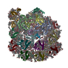
- PDB-9se7: Structure of Cytochrome C6 bound Photosystem I from Chlamydomonas... -
+ Open data

- Basic information
| Entry | Database: PDB / ID: 9se7 | ||||||||||||||||||||||||
|---|---|---|---|---|---|---|---|---|---|---|---|---|---|---|---|---|---|---|---|---|---|---|---|---|---|
| Title | Structure of Cytochrome C6 bound Photosystem I from Chlamydomonas reinhardtii at 2.07 A resolution | ||||||||||||||||||||||||
 Components Components |
| ||||||||||||||||||||||||
 Keywords Keywords | PHOTOSYNTHESIS / Cytochrome C6 / Photosystem I / Chlamydomonas reinhardtii / CryoEM | ||||||||||||||||||||||||
| Function / homology |  Function and homology information Function and homology informationchloroplast thylakoid lumen / photosystem I reaction center / photosystem I / photosynthetic electron transport in photosystem I / photosystem I / chlorophyll binding / chloroplast thylakoid membrane / photosynthesis / 4 iron, 4 sulfur cluster binding / electron transfer activity ...chloroplast thylakoid lumen / photosystem I reaction center / photosystem I / photosynthetic electron transport in photosystem I / photosystem I / chlorophyll binding / chloroplast thylakoid membrane / photosynthesis / 4 iron, 4 sulfur cluster binding / electron transfer activity / oxidoreductase activity / iron ion binding / heme binding / magnesium ion binding / metal ion binding Similarity search - Function | ||||||||||||||||||||||||
| Biological species |   Chlamydomonas reinhardtii (plant) Chlamydomonas reinhardtii (plant) | ||||||||||||||||||||||||
| Method | ELECTRON MICROSCOPY / single particle reconstruction / cryo EM / Resolution: 2.06 Å | ||||||||||||||||||||||||
 Authors Authors | Mahapatra, G.P. / Schuller, J.M. | ||||||||||||||||||||||||
| Funding support |  Germany, 1items Germany, 1items
| ||||||||||||||||||||||||
 Citation Citation | Journal: Nat Commun / Year: 2026 Title: Cryo-EM structure of Chlamydomonas reinhardtii Photosystem I complexed with cytochrome c. Authors: Yu Ogawa / Gyana Prakash Mahapatra / Yuval Milrad / Michelle Schimpf / Genji Kurisu / Michael Hippler / Jan Michael Schuller /  Abstract: Photosynthetic electron transfer relies on small soluble carriers that shuttle electrons between the cytochrome b₆f complex and Photosystem I (PSI). While copper-containing plastocyanin (Pc) serves ...Photosynthetic electron transfer relies on small soluble carriers that shuttle electrons between the cytochrome b₆f complex and Photosystem I (PSI). While copper-containing plastocyanin (Pc) serves this role in plants, the heme protein cytochrome c₆ (Cyt c₆) is also employed in algae and cyanobacteria. Here, we present a cryo-electron microscopy structure of a Cyt c₆:PSI complex from Chlamydomonas reinhardtii. We observe that the heme group of Cyt c₆ is positioned ~11 Å away from P700, stabilized by extensive contacts involving a N-terminal helix-loop-helix motif of PSAF, characteristic of eukaryotic PSI. Notably, the algal Cyt c₆ also retains an arginine residue (R66) which is crucial for cyanobacterial donor:PSI reactions. Our structure reveals the previously uncharacterized interactions involving this residue; it can form a putative electrostatic contact with PsaB-D623 while also contributing to a tri-planar π(cation)-π interactions with adjacent residues. Our findings provide a structural framework for understanding the mechanism and evolution of donor:PSI interactions. | ||||||||||||||||||||||||
| History |
|
- Structure visualization
| Structure viewer | Molecule: MolmilJmol/JSmol |
|---|
- Downloads & links
-Download
| PDBx/mmCIF format | 9se7.cif.gz | 768.7 KB | Display | PDBx/mmCIF format |
|---|---|---|---|---|
| PDB format | pdb9se7.ent.gz | 535.1 KB | Display | PDB format |
| PDBx/mmJSON format | 9se7.json.gz | Tree view | PDBx/mmJSON format | |
| Others |  Other downloads Other downloads |
-Validation report
| Arichive directory | https://data.pdbj.org/pub/pdb/validation_reports/se/9se7ftp://data.pdbj.org/pub/pdb/validation_reports/se/9se7 | HTTPS FTP |
|---|
-Related structure data
| Related structure data |  54804MC  9se6C C: citing same article ( M: map data used to model this data |
|---|---|
| Similar structure data |

-Links
 PDBj
PDBj


- Assembly
| Deposited unit | 
|
|---|---|
| 1 |
|
-Components
-Photosystem I P700 chlorophyll a apoprotein ... , 2 types, 2 molecules AB
| #1: Protein | Mass: 82193.023 Da / Num. of mol.: 1 / Source method: isolated from a natural source / Source: (natural) Chlamydomonas reinhardtii (plant) / References: UniProt: P12154, photosystem I |
|---|---|
| #2: Protein | Mass: 81981.992 Da / Num. of mol.: 1 / Source method: isolated from a natural source / Source: (natural) Chlamydomonas reinhardtii (plant) / References: UniProt: P09144, photosystem I |
-Protein , 2 types, 2 molecules CT
| #3: Protein | Mass: 8738.130 Da / Num. of mol.: 1 / Source method: isolated from a natural source / Source: (natural) Chlamydomonas reinhardtii (plant) / References: UniProt: Q00914, photosystem I |
|---|---|
| #8: Protein | Mass: 9647.821 Da / Num. of mol.: 1 / Source method: isolated from a natural source / Source: (natural) Chlamydomonas reinhardtii (plant) / References: UniProt: P08197 |
-Photosystem I reaction center subunit ... , 6 types, 6 molecules DEIJFL
| #4: Protein | Mass: 16123.686 Da / Num. of mol.: 1 / Source method: isolated from a natural source / Source: (natural) Chlamydomonas reinhardtii (plant) / References: UniProt: Q39615 |
|---|---|
| #5: Protein | Mass: 7151.057 Da / Num. of mol.: 1 / Source method: isolated from a natural source / Source: (natural) Chlamydomonas reinhardtii (plant) / References: UniProt: P12352 |
| #6: Protein/peptide | Mass: 3970.731 Da / Num. of mol.: 1 / Source method: isolated from a natural source / Source: (natural) Chlamydomonas reinhardtii (plant) / References: UniProt: A8IFG7 |
| #7: Protein/peptide | Mass: 4629.373 Da / Num. of mol.: 1 / Source method: isolated from a natural source / Source: (natural) Chlamydomonas reinhardtii (plant) / References: UniProt: P59777 |
| #9: Protein | Mass: 17957.773 Da / Num. of mol.: 1 / Source method: isolated from a natural source / Source: (natural) Chlamydomonas reinhardtii (plant) / References: UniProt: P12356 |
| #10: Protein | Mass: 14130.248 Da / Num. of mol.: 1 / Source method: isolated from a natural source / Source: (natural) Chlamydomonas reinhardtii (plant) / References: UniProt: A8IL32 |
-Sugars , 2 types, 4 molecules 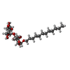

| #16: Sugar |  Type: D-saccharide / Mass: 510.615 Da / Num. of mol.: 3 / Source method: obtained synthetically / Formula: C24H46O11 / Comment: detergent*YM Type: D-saccharide / Mass: 510.615 Da / Num. of mol.: 3 / Source method: obtained synthetically / Formula: C24H46O11 / Comment: detergent*YM#18: Sugar | ChemComp-DGD / |  Type: saccharide / Mass: 949.299 Da / Num. of mol.: 1 / Source method: obtained synthetically / Formula: C51H96O15 Type: saccharide / Mass: 949.299 Da / Num. of mol.: 1 / Source method: obtained synthetically / Formula: C51H96O15 |
|---|
-Non-polymers , 9 types, 374 molecules 


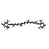


| #11: Chemical | ChemComp-CLA /  Mass: 893.489 Da / Num. of mol.: 91 / Source method: obtained synthetically / Formula: C55H72MgN4O5 / Feature type: SUBJECT OF INVESTIGATION Mass: 893.489 Da / Num. of mol.: 91 / Source method: obtained synthetically / Formula: C55H72MgN4O5 / Feature type: SUBJECT OF INVESTIGATION#12: Chemical |  Mass: 450.696 Da / Num. of mol.: 2 / Source method: obtained synthetically / Formula: C31H46O2 / Feature type: SUBJECT OF INVESTIGATION Mass: 450.696 Da / Num. of mol.: 2 / Source method: obtained synthetically / Formula: C31H46O2 / Feature type: SUBJECT OF INVESTIGATION#13: Chemical |  Mass: 722.970 Da / Num. of mol.: 3 / Source method: obtained synthetically / Formula: C38H75O10P / Comment: phospholipid*YM Mass: 722.970 Da / Num. of mol.: 3 / Source method: obtained synthetically / Formula: C38H75O10P / Comment: phospholipid*YM#14: Chemical | ChemComp-BCR /  Mass: 536.873 Da / Num. of mol.: 15 / Source method: obtained synthetically / Formula: C40H56 / Feature type: SUBJECT OF INVESTIGATION Mass: 536.873 Da / Num. of mol.: 15 / Source method: obtained synthetically / Formula: C40H56 / Feature type: SUBJECT OF INVESTIGATION#15: Chemical |  Mass: 351.640 Da / Num. of mol.: 3 / Source method: obtained synthetically / Formula: Fe4S4 / Feature type: SUBJECT OF INVESTIGATION Mass: 351.640 Da / Num. of mol.: 3 / Source method: obtained synthetically / Formula: Fe4S4 / Feature type: SUBJECT OF INVESTIGATION#17: Chemical | ChemComp-LMG /  Mass: 787.158 Da / Num. of mol.: 5 / Source method: obtained synthetically / Formula: C45H86O10 Mass: 787.158 Da / Num. of mol.: 5 / Source method: obtained synthetically / Formula: C45H86O10#19: Chemical |  Mass: 568.871 Da / Num. of mol.: 2 / Source method: obtained synthetically / Formula: C40H56O2 / Feature type: SUBJECT OF INVESTIGATION Mass: 568.871 Da / Num. of mol.: 2 / Source method: obtained synthetically / Formula: C40H56O2 / Feature type: SUBJECT OF INVESTIGATION#20: Chemical | ChemComp-HEC / |  Mass: 618.503 Da / Num. of mol.: 1 / Source method: obtained synthetically / Formula: C34H34FeN4O4 / Feature type: SUBJECT OF INVESTIGATION Mass: 618.503 Da / Num. of mol.: 1 / Source method: obtained synthetically / Formula: C34H34FeN4O4 / Feature type: SUBJECT OF INVESTIGATION#21: Water | ChemComp-HOH / | Mass: 18.015 Da / Num. of mol.: 252 / Source method: isolated from a natural source / Formula: H2O |
|---|
-Details
| Has ligand of interest | Y |
|---|---|
| Has protein modification | Y |
-Experimental details
-Experiment
| Experiment | Method: ELECTRON MICROSCOPY |
|---|---|
| EM experiment | Aggregation state: PARTICLE / 3D reconstruction method: single particle reconstruction |
- Sample preparation
| Component | Name: Cytochrome C6 bound Photosystem I / Type: COMPLEX / Entity ID: #1-#10 / Source: NATURAL |
|---|---|
| Source (natural) | Organism: Chlamydomonas reinhardtii (plant) |
| Buffer solution | pH: 7.5 / Details: 5mM HEPES, pH 7.5, 0.02% alphaDDM |
| Specimen | Conc.: 1.5 mg/ml / Embedding applied: NO / Shadowing applied: NO / Staining applied: NO / Vitrification applied: YES |
| Specimen support | Grid material: COPPER / Grid mesh size: 200 divisions/in. / Grid type: Quantifoil R2/1 |
| Vitrification | Instrument: FEI VITROBOT MARK IV / Cryogen name: ETHANE / Humidity: 100 % / Chamber temperature: 277.15 K |
- Electron microscopy imaging
| Experimental equipment |  Model: Titan Krios / Image courtesy: FEI Company |
|---|---|
| Microscopy | Model: TFS KRIOS |
| Electron gun | Electron source:  FIELD EMISSION GUN / Accelerating voltage: 300 kV / Illumination mode: OTHER FIELD EMISSION GUN / Accelerating voltage: 300 kV / Illumination mode: OTHER |
| Electron lens | Mode: OTHER / Nominal defocus max: 2000 nm / Nominal defocus min: 800 nm / Cs: 2.7 mm |
| Image recording | Average exposure time: 1.9 sec. / Electron dose: 50 e/Å2 / Film or detector model: FEI FALCON IV (4k x 4k) / Num. of grids imaged: 4 |
- Processing
| EM software |
| ||||||||||||||||||||||||||||||||||||
|---|---|---|---|---|---|---|---|---|---|---|---|---|---|---|---|---|---|---|---|---|---|---|---|---|---|---|---|---|---|---|---|---|---|---|---|---|---|
| CTF correction | Type: PHASE FLIPPING AND AMPLITUDE CORRECTION | ||||||||||||||||||||||||||||||||||||
| Particle selection | Num. of particles selected: 30 | ||||||||||||||||||||||||||||||||||||
| 3D reconstruction | Resolution: 2.06 Å / Resolution method: FSC 0.143 CUT-OFF / Num. of particles: 53904 / Algorithm: FOURIER SPACE / Num. of class averages: 1 / Symmetry type: POINT | ||||||||||||||||||||||||||||||||||||
| Atomic model building | Protocol: RIGID BODY FIT / Space: REAL | ||||||||||||||||||||||||||||||||||||
| Atomic model building |
| ||||||||||||||||||||||||||||||||||||
| Refinement | Cross valid method: NONE Stereochemistry target values: GeoStd + Monomer Library + CDL v1.2 | ||||||||||||||||||||||||||||||||||||
| Displacement parameters | Biso mean: 51.92 Å2 | ||||||||||||||||||||||||||||||||||||
| Refine LS restraints |
|