National Institutes of Health/National Heart, Lung, and Blood Institute (NIH/NHLBI)
R35 HL135823
米国
National Institutes of Health/National Heart, Lung, and Blood Institute (NIH/NHLBI)
R35 HL171334
米国
National Institutes of Health/Office of the Director
S10OD030275
米国
National Institutes of Health/National Institute of General Medical Sciences (NIH/NIGMS)
R24 GM145965
米国
American Heart Association
N028347
米国
引用
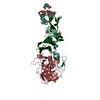
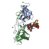
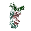
ジャーナル: Blood / 年: 2025 タイトル: Cryo-EM structure of the tissue factor/factor VIIa complex with a factor X mimetic reveals a novel allosteric mechanism. 著者: Josepha C Sedzro / Amanda L Photenhauer / Fabienne Birkle / Katarina Meze / Alex Mortenson / Cade Duckworth / Po-Chao Wen / Sarah Kearns / Michael A Cianfrocco / Emad Tajkhorshid / Melanie D ...著者: Josepha C Sedzro / Amanda L Photenhauer / Fabienne Birkle / Katarina Meze / Alex Mortenson / Cade Duckworth / Po-Chao Wen / Sarah Kearns / Michael A Cianfrocco / Emad Tajkhorshid / Melanie D Ohi / James H Morrissey / 要旨: Blood clotting is triggered in hemostasis and thrombosis when the membrane-bound tissue factor (TF)/factor VIIa (FVIIa) complex activates factor X (FX). There are no structures of TF/FVIIa on ...Blood clotting is triggered in hemostasis and thrombosis when the membrane-bound tissue factor (TF)/factor VIIa (FVIIa) complex activates factor X (FX). There are no structures of TF/FVIIa on membranes, with or without FX. Using cryo-EM to address this gap, we assembled TF/FVIIa complexes on nanoscale membrane bilayers (nanodiscs), bound to XK1 and an antibody fragment. XK1 is a FX mimetic whose protease domain is replaced by the first Kunitz-type (K1) domain of tissue factor pathway inhibitor, while 10H10 is a non-inhibitory, anti-TF antibody. We determined a cryo-EM structure of a TF/FVIIa/XK1/10H10/nanodisc complex with a resolution of 3.7 Å, allowing us to model all the protein backbones. TF/FVIIa extends perpendicularly from the membrane, interacting with a "handle shaped" XK1 at two locations: the K1 domain docks into FVIIa's active site, while the γ-carboxyglutamate-rich (GLA) domain binds to TF's substrate-binding exosite. The FX and FVIIa GLA domains also contact each other and the membrane surface. Except for a minor contact between the first epidermal growth factor (EGF)-like domain of XK1 and TF, the rest of the FX light chain does not interact with TF/FVIIa. The structure reveals a previously unrecognized, membrane-dependent allosteric activation mechanism between FVIIa and TF where a serine-rich loop in TF that partially obscures the TF exosite must undergo a shift to allow access of the FX GLA domain to its final binding location on the membrane-bound TF/FVIIa complex. This mechanism also provides a novel explanation for the otherwise puzzling phenomenon of TF encryption/decryption on cell surfaces.
V: Factor VII light chain X: Factor X light chain S: Coagulation factor VII Heavy Chain T: Tissue factor,Maltose/maltodextrin-binding periplasmic protein K: Tissue factor pathway inhibitor H: Human 10H10 antibody Fab heavy chain L: Human 10H10 antibody Fab light chain ヘテロ分子
解像度: 3.7 Å / 解像度の算出法: FSC 0.143 CUT-OFF / 粒子像の数: 20107 / 対称性のタイプ: POINT
原子モデル構築
プロトコル: FLEXIBLE FIT / 空間: REAL / Target criteria: cross-correlation 詳細: Crystal structures were rigid-body fit into the density map and model optimization was then carried out with Phenix real-space refine. Manual refinement in Coot was performed to ensure that ...詳細: Crystal structures were rigid-body fit into the density map and model optimization was then carried out with Phenix real-space refine. Manual refinement in Coot was performed to ensure that the backbone traces followed the density in regions where the map was high enough resolution to trace the backbone. Secondary structure restraints were used to ensure that alpha-helices and beta-sheets did not deviate far from their expected geometry. A final check of MolProbity and cross correlation was done to ensure model quality.
原子モデル構築
3D fitting-ID: 1 / Source name: PDB / タイプ: experimental model
 ムービー
ムービー コントローラー


 データを開く
 基本情報
基本情報 要素
要素 キーワード
キーワード 機能・相同性情報
 Homo sapiens (ヒト)

 データ登録者
 米国, 5件
  引用
引用 構造の表示
構造の表示 ダウンロードとリンク
 その他のダウンロード


 PDBj
PDBj


 集合体
集合体


 タイプ: D-saccharide, beta linking / 分子量: 180.156 Da / 分子数: 1 / 由来タイプ: 合成 / 式: C6H12O6
 タイプ: L-saccharide, alpha linking / 分子量: 164.156 Da / 分子数: 1 / 由来タイプ: 合成 / 式: C6H12O5

 分子量: 24.305 Da / 分子数: 4 / 由来タイプ: 合成 / 式: Mg
 分子量: 40.078 Da / 分子数: 12 / 由来タイプ: 合成 / 式: Ca
 試料調製
試料調製 電子顕微鏡撮影
 FIELD EMISSION GUN / 加速電圧: 300 kV / 照射モード: FLOOD BEAM
 解析
解析