 Movie
Movie Controller

[English] 日本語
 Yorodumi
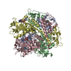
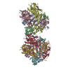
- PDB-9nlg: CBASS Pseudomonas syringae Cap5 tetramer with 3'2'-c-GAMP cyclic ... -
+ Open data

- Basic information
| Entry | Database: PDB / ID: 9nlg | ||||||
|---|---|---|---|---|---|---|---|
| Title | CBASS Pseudomonas syringae Cap5 tetramer with 3'2'-c-GAMP cyclic dinucleotide ligand (His56Ala mutant without Mg2+ ions) | ||||||
 Components Components | HNH endonuclease | ||||||
 Keywords Keywords | IMMUNE SYSTEM / Bacterial immunity / CBASS / cyclic dinucleotide / Cap5 effector DNA endonuclease / viral defense / DNA | ||||||
| Function / homology | HNH endonuclease / SMODS-associated and fused to various effectors / SMODS-associated and fused to various effectors sensor domain / HNH nuclease / metal ion binding / 3'2'-cGAMP / HNH endonuclease Function and homology information Function and homology information | ||||||
| Biological species |  Pseudomonas syringae (bacteria) Pseudomonas syringae (bacteria) | ||||||
| Method |  X-RAY DIFFRACTION / SYNCHROTRON / MOLECULAR REPLACEMENT / Resolution: 1.64 Å X-RAY DIFFRACTION / SYNCHROTRON / MOLECULAR REPLACEMENT / Resolution: 1.64 Å | ||||||
 Authors Authors | Rechkoblit, O. / Aggarwal, A.K. | ||||||
| Funding support |  United States, 1items United States, 1items
| ||||||
 Citation Citation | Journal: Nat Commun / Year: 2025 Title: Mechanism of DNA degradation by CBASS Cap5 endonuclease immune effector. Authors: Rechkoblit, O. / Sciaky, D. / Ni, M. / Li, Y. / Kottur, J. / Fang, G. / Aggarwal, A.K. | ||||||
| History |
|
- Structure visualization
| Structure viewer | Molecule: MolmilJmol/JSmol |
|---|
- Downloads & links
-Download
| PDBx/mmCIF format | 9nlg.cif.gz | 700.8 KB | Display | PDBx/mmCIF format |
|---|---|---|---|---|
| PDB format | pdb9nlg.ent.gz | 468.8 KB | Display | PDB format |
| PDBx/mmJSON format | 9nlg.json.gz | Tree view | PDBx/mmJSON format | |
| Others |  Other downloads Other downloads |
-Validation report
| Arichive directory | https://data.pdbj.org/pub/pdb/validation_reports/nl/9nlgftp://data.pdbj.org/pub/pdb/validation_reports/nl/9nlg | HTTPS FTP |
|---|
-Related structure data


-Links
 PDBj
PDBj


- Assembly
| Deposited unit | 
| ||||||||||||
|---|---|---|---|---|---|---|---|---|---|---|---|---|---|
| 1 |
| ||||||||||||
| Unit cell |
|
-Components
| #1: Protein | Mass: 42702.168 Da / Num. of mol.: 4 / Mutation: H56A Source method: isolated from a genetically manipulated source Source: (gene. exp.) Pseudomonas syringae (bacteria) / Production host: #2: Chemical | ChemComp-ZN /   Mass: 65.409 Da / Num. of mol.: 4 / Source method: obtained synthetically / Formula: Zn Mass: 65.409 Da / Num. of mol.: 4 / Source method: obtained synthetically / Formula: Zn#3: Chemical | ChemComp-4UR / 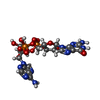  Mass: 674.411 Da / Num. of mol.: 4 / Source method: obtained synthetically / Formula: C20H24N10O13P2 / Feature type: SUBJECT OF INVESTIGATION Mass: 674.411 Da / Num. of mol.: 4 / Source method: obtained synthetically / Formula: C20H24N10O13P2 / Feature type: SUBJECT OF INVESTIGATION#4: Chemical | ChemComp-GOL / |   Mass: 92.094 Da / Num. of mol.: 1 / Source method: obtained synthetically / Formula: C3H8O3 Mass: 92.094 Da / Num. of mol.: 1 / Source method: obtained synthetically / Formula: C3H8O3#5: Water | ChemComp-HOH / |  Mass: 18.015 Da / Num. of mol.: 1500 / Source method: isolated from a natural source / Formula: H2O Mass: 18.015 Da / Num. of mol.: 1500 / Source method: isolated from a natural source / Formula: H2OHas ligand of interest | Y | Has protein modification | N | |
|---|
-Experimental details
-Experiment
| Experiment | Method: X-RAY DIFFRACTION / Number of used crystals: 1 |
|---|
- Sample preparation
| Crystal | Density Matthews: 2.28 Å3/Da / Density % sol: 46.02 % |
|---|---|
| Crystal grow | Temperature: 293 K / Method: vapor diffusion, hanging drop / pH: 9 / Details: 10 mM Trisodiumcitrate pH 9.0, 28% PEG6000 |
-Data collection
| Diffraction | Mean temperature: 100 K / Serial crystal experiment: N |
|---|---|
| Diffraction source | Source: SYNCHROTRON / Site: NSLS-II / Beamline: 17-ID-2 / Wavelength: 0.979338 Å |
| Detector | Type: DECTRIS EIGER2 S 16M / Detector: PIXEL / Date: Jul 14, 2023 |
| Radiation | Protocol: SINGLE WAVELENGTH / Monochromatic (M) / Laue (L): M / Scattering type: x-ray |
| Radiation wavelength | Wavelength: 0.979338 Å / Relative weight: 1 |
| Reflection | Resolution: 1.64→69.054 Å / Num. obs: 173836 / % possible obs: 94.7 % / Redundancy: 2.8 % / Biso Wilson estimate: 18.65 Å2 / CC1/2: 0.998 / Rmerge(I) obs: 0.055 / Net I/σ(I): 10.3 |
| Reflection shell | Resolution: 1.64→1.683 Å / Rmerge(I) obs: 0.547 / Num. unique obs: 8697 / CC1/2: 0.687 |
- Processing
| Software |
| |||||||||||||||||||||||||||||||||||||||||||||||||||||||||||||||||||||||||||||||||||||||||||||||||||||||||||||||||||||||||||||||||||||||||||||||||||||||||||||||||||||||||||||||||||||||||||||||||||||||||||||||||||||||||
|---|---|---|---|---|---|---|---|---|---|---|---|---|---|---|---|---|---|---|---|---|---|---|---|---|---|---|---|---|---|---|---|---|---|---|---|---|---|---|---|---|---|---|---|---|---|---|---|---|---|---|---|---|---|---|---|---|---|---|---|---|---|---|---|---|---|---|---|---|---|---|---|---|---|---|---|---|---|---|---|---|---|---|---|---|---|---|---|---|---|---|---|---|---|---|---|---|---|---|---|---|---|---|---|---|---|---|---|---|---|---|---|---|---|---|---|---|---|---|---|---|---|---|---|---|---|---|---|---|---|---|---|---|---|---|---|---|---|---|---|---|---|---|---|---|---|---|---|---|---|---|---|---|---|---|---|---|---|---|---|---|---|---|---|---|---|---|---|---|---|---|---|---|---|---|---|---|---|---|---|---|---|---|---|---|---|---|---|---|---|---|---|---|---|---|---|---|---|---|---|---|---|---|---|---|---|---|---|---|---|---|---|---|---|---|---|---|---|---|
| Refinement | Method to determine structure: MOLECULAR REPLACEMENT / Resolution: 1.64→45.87 Å / SU ML: 0.1854 / Cross valid method: FREE R-VALUE / σ(F): 1.98 / Phase error: 17.2105 Stereochemistry target values: GeoStd + Monomer Library + CDL v1.2
| |||||||||||||||||||||||||||||||||||||||||||||||||||||||||||||||||||||||||||||||||||||||||||||||||||||||||||||||||||||||||||||||||||||||||||||||||||||||||||||||||||||||||||||||||||||||||||||||||||||||||||||||||||||||||
| Solvent computation | Shrinkage radii: 0.9 Å / VDW probe radii: 1.1 Å / Solvent model: FLAT BULK SOLVENT MODEL | |||||||||||||||||||||||||||||||||||||||||||||||||||||||||||||||||||||||||||||||||||||||||||||||||||||||||||||||||||||||||||||||||||||||||||||||||||||||||||||||||||||||||||||||||||||||||||||||||||||||||||||||||||||||||
| Displacement parameters | Biso mean: 23.85 Å2 | |||||||||||||||||||||||||||||||||||||||||||||||||||||||||||||||||||||||||||||||||||||||||||||||||||||||||||||||||||||||||||||||||||||||||||||||||||||||||||||||||||||||||||||||||||||||||||||||||||||||||||||||||||||||||
| Refinement step | Cycle: LAST / Resolution: 1.64→45.87 Å
| |||||||||||||||||||||||||||||||||||||||||||||||||||||||||||||||||||||||||||||||||||||||||||||||||||||||||||||||||||||||||||||||||||||||||||||||||||||||||||||||||||||||||||||||||||||||||||||||||||||||||||||||||||||||||
| Refine LS restraints |
| |||||||||||||||||||||||||||||||||||||||||||||||||||||||||||||||||||||||||||||||||||||||||||||||||||||||||||||||||||||||||||||||||||||||||||||||||||||||||||||||||||||||||||||||||||||||||||||||||||||||||||||||||||||||||
| LS refinement shell |
|
