- PDB-9mot: Cryo-EM structure of factor Va bound to activated protein C -
+
Open data
ID or keywords:
Loading...
-
Basic information
Entry
Database: PDB / ID: 9mot
Title
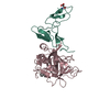
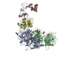
Cryo-EM structure of factor Va bound to activated protein C
Components
Coagulation factor Va heavy chain
Coagulation factor Va light chain
Vitamin K-dependent protein C heavy chain
Keywords
BLOOD CLOTTING / Coagulation / Activated Factor V / Activated Protein C
Function / homology
Function and homology information
activated protein C (thrombin-activated peptidase) / positive regulation of establishment of endothelial barrier / negative regulation of coagulation / response to vitamin K / platelet alpha granule / Cargo concentration in the ER / COPII-coated ER to Golgi transport vesicle / COPII-mediated vesicle transport / blood circulation / negative regulation of blood coagulation ...activated protein C (thrombin-activated peptidase) / positive regulation of establishment of endothelial barrier / negative regulation of coagulation / response to vitamin K / platelet alpha granule / Cargo concentration in the ER / COPII-coated ER to Golgi transport vesicle / COPII-mediated vesicle transport / blood circulation / negative regulation of blood coagulation / Transport of gamma-carboxylated protein precursors from the endoplasmic reticulum to the Golgi apparatus / : / Gamma-carboxylation of protein precursors / Removal of aminoterminal propeptides from gamma-carboxylated proteins / : / endoplasmic reticulum-Golgi intermediate compartment membrane / platelet alpha granule lumen / Cell surface interactions at the vascular wall / Post-translational protein phosphorylation / Golgi lumen / negative regulation of inflammatory response / Regulation of Insulin-like Growth Factor (IGF) transport and uptake by Insulin-like Growth Factor Binding Proteins (IGFBPs) / blood coagulation / Platelet degranulation / extracellular vesicle / endoplasmic reticulum lumen / copper ion binding / serine-type endopeptidase activity / calcium ion binding / negative regulation of apoptotic process / Golgi apparatus / endoplasmic reticulum / proteolysis / : / extracellular region / membrane / plasma membrane Similarity search - Function
National Institutes of Health/National Heart, Lung, and Blood Institute (NIH/NHLBI)
HL049413, HL139554 and HL147821
United States
Childrens Discovery Institute of Washington University and St. Louis Childrens Hospital
CDI-CORE-2015-505 and CDI-CORE-2019-813
United States
The Foundation for Barnes-Jewish Hospital
3770
United States
National Institutes of Health/National Institute of Diabetes and Digestive and Kidney Disease (NIH/NIDDK)
DK020579
United States
National Institutes of Health/National Cancer Institute (NIH/NCI)
CA091842
United States
Citation
Journal: Blood / Year: 2025 Title: Cryo-EM structure of coagulation factor Va bound to activated protein C. Authors: Bassem M Mohammed / Katherine Basore / Enrico Di Cera / Abstract: Coagulation factor Va (FVa) is the cofactor component of the prothrombinase complex required for rapid generation of thrombin from prothrombin in the penultimate step of the coagulation cascade. In ...Coagulation factor Va (FVa) is the cofactor component of the prothrombinase complex required for rapid generation of thrombin from prothrombin in the penultimate step of the coagulation cascade. In addition, FVa is a target for proteolytic inactivation by activated protein C (APC). Like other protein-protein interactions in the coagulation cascade, the FVa-APC interaction has long posed a challenge to structural biology and its molecular underpinnings remain unknown. A recent cryogenic electron microscopy (cryo-EM) structure of FVa has revealed the arrangement of its A1-A2-A3-C1-C2 domains and the environment of the sites of APC cleavage at R306 and R506. Here, we report the cryo-EM structure of the FVa-APC complex at 3.15 Å resolution in which the protease domain of APC engages R506 in the A2 domain of FVa through electrostatic interactions between positively charged residues in the 30-loop and 70-loop of APC and an electronegative surface of FVa. The auxiliary γ-carboxyglutamic acid and epidermal growth factor domains of APC are highly dynamic and point to solvent, without making contacts with FVa. Binding of APC displaces a large portion of the A2 domain of FVa and projects the 654VKCIPDDDEDSYEIFEP670 segment as a "latch," or exosite ligand, over the 70-loop of the enzyme. The latch induces a large conformational change of the autolysis loop of APC, which in turn promotes docking of R506 into the primary specificity pocket. The cryo-EM structure of the FVa-APC complex validates the bulk of existing biochemical data and offers molecular context for a key regulatory interaction of the coagulation cascade.
Imaging-ID: 1 / Film or detector model: FEI FALCON IV (4k x 4k) / Num. of grids imaged: 1
ID
Electron dose (e/Å2)
Detector mode
Num. of real images
Details (eV)
1
51.86
COUNTING
3193
30degreestilt
2
46.6
600
3
46.89
2655
30degreestilt
4
47.19
2379
5
46.89
420
6
52.8
3210
Image scans
Width
Height
Movie frames/image
ID
Image recording-ID
Entry-ID
4096
4096
50
1
1
9MOT
4096
4096
2
2
9MOT
4096
4096
3
3
9MOT
4096
4096
4
4
9MOT
4096
4096
5
5
9MOT
4096
4096
6
6
9MOT
-
Processing
EM software
ID
Name
Version
Category
1
cryoSPARC
4.6.2
particleselection
2
EPU
imageacquisition
4
cryoSPARC
4.6.2
CTFcorrection
7
UCSF ChimeraX
1.9
modelfitting
12
cryoSPARC
4.6.2
3Dreconstruction
13
PHENIX
1.21.2-5419
modelrefinement
14
Coot
0.9.8.95
modelrefinement
CTF correction
Type: PHASE FLIPPING AND AMPLITUDE CORRECTION
Symmetry
Point symmetry: C1 (asymmetric)
3D reconstruction
Resolution: 3.15 Å / Resolution method: FSC 0.143 CUT-OFF / Num. of particles: 384239 / Algorithm: BACK PROJECTION Details: 3D flex with custom mesh was used for the reconstruction Num. of class averages: 1 / Symmetry type: POINT
In the structure databanks used in Yorodumi, some data are registered as the other names, "COVID-19 virus" and "2019-nCoV". Here are the details of the virus and the list of structure data.
Jan 31, 2019. EMDB accession codes are about to change! (news from PDBe EMDB page)
EMDB accession codes are about to change! (news from PDBe EMDB page)
The allocation of 4 digits for EMDB accession codes will soon come to an end. Whilst these codes will remain in use, new EMDB accession codes will include an additional digit and will expand incrementally as the available range of codes is exhausted. The current 4-digit format prefixed with “EMD-” (i.e. EMD-XXXX) will advance to a 5-digit format (i.e. EMD-XXXXX), and so on. It is currently estimated that the 4-digit codes will be depleted around Spring 2019, at which point the 5-digit format will come into force.
The EM Navigator/Yorodumi systems omit the EMD- prefix.
Related info.:Q: What is EMD? / ID/Accession-code notation in Yorodumi/EM Navigator
Yorodumi is a browser for structure data from EMDB, PDB, SASBDB, etc.
This page is also the successor to EM Navigator detail page, and also detail information page/front-end page for Omokage search.
The word "yorodu" (or yorozu) is an old Japanese word meaning "ten thousand". "mi" (miru) is to see.
Related info.:EMDB / PDB / SASBDB / Comparison of 3 databanks / Yorodumi Search / Aug 31, 2016. New EM Navigator & Yorodumi / Yorodumi Papers / Jmol/JSmol / Function and homology information / Changes in new EM Navigator and Yorodumi
 Movie
Movie Controller


 Open data
 Basic information
 Components
 Keywords
 Function and homology information
 Homo sapiens (human)
 Authors
 United States, 5items
  Citation
 Structure visualization
 Downloads & links
 Other downloads


 PDBj
PDBj


 Assembly
 Mesocricetus auratus (golden hamster) / References: UniProt: P04070
 Type: D-saccharide, beta linking / Mass: 221.208 Da / Num. of mol.: 8 / Source method: isolated from a natural source / Formula: C8H15NO6 / Feature type: SUBJECT OF INVESTIGATION
 Sample preparation
 Electron microscopy imaging
 FIELD EMISSION GUN / Accelerating voltage: 300 kV / Illumination mode: FLOOD BEAM
 Processing