 Movie
Movie Controller

[English] 日本語
 Yorodumi
- PDB-8tn9: Structural architecture of the acidic region of the B domain of c... -
+ Open data

- Basic information
| Entry | Database: PDB / ID: 8tn9 | ||||||||||||||||||
|---|---|---|---|---|---|---|---|---|---|---|---|---|---|---|---|---|---|---|---|
| Title | Structural architecture of the acidic region of the B domain of coagulation factor V | ||||||||||||||||||
 Components Components | Coagulation factor V | ||||||||||||||||||
 Keywords Keywords | BLOOD CLOTTING / Coagulation / Pro-cofactor / Factor V / B Domain / Acidic Region | ||||||||||||||||||
| Function / homology |  Function and homology information Function and homology informationresponse to vitamin K / platelet alpha granule / Cargo concentration in the ER / COPII-coated ER to Golgi transport vesicle / COPII-mediated vesicle transport / blood circulation / : / endoplasmic reticulum-Golgi intermediate compartment membrane / platelet alpha granule lumen / Post-translational protein phosphorylation ...response to vitamin K / platelet alpha granule / Cargo concentration in the ER / COPII-coated ER to Golgi transport vesicle / COPII-mediated vesicle transport / blood circulation / : / endoplasmic reticulum-Golgi intermediate compartment membrane / platelet alpha granule lumen / Post-translational protein phosphorylation / Regulation of Insulin-like Growth Factor (IGF) transport and uptake by Insulin-like Growth Factor Binding Proteins (IGFBPs) / blood coagulation / Platelet degranulation / extracellular vesicle / endoplasmic reticulum lumen / copper ion binding / : / extracellular region / membrane / plasma membrane Similarity search - Function | ||||||||||||||||||
| Biological species |  Homo sapiens (human) Homo sapiens (human) | ||||||||||||||||||
| Method | ELECTRON MICROSCOPY / single particle reconstruction / cryo EM / Resolution: 3.05 Å | ||||||||||||||||||
 Authors Authors | Mohammed, B.M. / Basore, K. / Summers, B. / Pelc, L.A. / Di Cera, E. | ||||||||||||||||||
| Funding support |  United States, 5items United States, 5items
| ||||||||||||||||||
 Citation Citation | Journal: J Thromb Haemost / Year: 2024 Title: Structural architecture of the acidic region of the B domain of coagulation factor V. Authors: Bassem M Mohammed / Katherine Basore / Brock Summers / Leslie A Pelc / Enrico Di Cera / Abstract: BACKGROUND: Coagulation factor (F)V features an A1-A2-B-A3-C1-C2 domain organization and functions as the inactive precursor of FVa, a component of the prothrombinase complex required for rapid ...BACKGROUND: Coagulation factor (F)V features an A1-A2-B-A3-C1-C2 domain organization and functions as the inactive precursor of FVa, a component of the prothrombinase complex required for rapid thrombin generation in the penultimate step of the coagulation cascade. An intramolecular interaction within the large B domain (residues 710-1545) involves the basic region (BR, residues 963-1008) and acidic region (AR, residues 1493-1537) and locks FV in its inactive state. However, structural information on this important regulatory interaction or on the separate architecture of the AR and BR remains elusive due to conformational disorder of the B domain. OBJECTIVES: To reveal the structure of the BR-AR interaction or of its separate components. METHODS: The structure of FV is solved by cryogenic electron microscopy. RESULTS: A new 3.05 Å resolution cryogenic electron microscopy structure of FV confirms the overall organization of the A and C domains but resolves the segment 1507 to 1545 within a largely ...RESULTS: A new 3.05 Å resolution cryogenic electron microscopy structure of FV confirms the overall organization of the A and C domains but resolves the segment 1507 to 1545 within a largely disordered B domain. The segment contains most of the AR and is organized as recently reported in FV short, a spliced variant of FV with a significantly shorter and less disordered B domain. CONCLUSION: The similar architecture of the AR in FV and FV short provides structural context for physiologically important interactions of this region with the BR in FV and with the basic C-terminal ...CONCLUSION: The similar architecture of the AR in FV and FV short provides structural context for physiologically important interactions of this region with the BR in FV and with the basic C-terminal end of tissue factor pathway inhibitor α in FV short. | ||||||||||||||||||
| History |
|
- Structure visualization
| Structure viewer | Molecule: MolmilJmol/JSmol |
|---|
- Downloads & links
-Download
| PDBx/mmCIF format | 8tn9.cif.gz | 303.8 KB | Display | PDBx/mmCIF format |
|---|---|---|---|---|
| PDB format | pdb8tn9.ent.gz | 230.7 KB | Display | PDB format |
| PDBx/mmJSON format | 8tn9.json.gz | Tree view | PDBx/mmJSON format | |
| Others |  Other downloads Other downloads |
-Validation report
| Arichive directory | https://data.pdbj.org/pub/pdb/validation_reports/tn/8tn9ftp://data.pdbj.org/pub/pdb/validation_reports/tn/8tn9 | HTTPS FTP |
|---|
-Related structure data
| Related structure data |  41411MC C: citing same article ( M: map data used to model this data |
|---|---|
| Similar structure data |


-Links
 PDBj
PDBj


- Assembly
| Deposited unit | 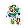
|
|---|---|
| 1 |
|
-Components
| #1: Protein | Mass: 248973.234 Da / Num. of mol.: 1 / Source method: isolated from a natural source / Source: (natural) Homo sapiens (human) / References: UniProt: P12259 | ||||
|---|---|---|---|---|---|
| #2: Sugar | ChemComp-NAG /   Type: D-saccharide, beta linking / Mass: 221.208 Da / Num. of mol.: 4 / Source method: obtained synthetically / Formula: C8H15NO6 / Feature type: SUBJECT OF INVESTIGATION Type: D-saccharide, beta linking / Mass: 221.208 Da / Num. of mol.: 4 / Source method: obtained synthetically / Formula: C8H15NO6 / Feature type: SUBJECT OF INVESTIGATIONHas ligand of interest | Y | Has protein modification | Y | |
-Experimental details
-Experiment
| Experiment | Method: ELECTRON MICROSCOPY |
|---|---|
| EM experiment | Aggregation state: PARTICLE / 3D reconstruction method: single particle reconstruction |
- Sample preparation
| Component | Name: Coagulation plasma Factor V / Type: TISSUE / Entity ID: #1 / Source: NATURAL |
|---|---|
| Source (natural) | Organism: Homo sapiens (human) / Tissue: Blood |
| Buffer solution | pH: 7.4 / Details: 20 mM HEPES, 150 mM NaCl, 5 mM CaCl2 |
| Specimen | Conc.: 0.2 mg/ml / Embedding applied: NO / Shadowing applied: NO / Staining applied: NO / Vitrification applied: YES / Details: 0.1 and 0.2 mg/mL |
| Specimen support | Grid material: COPPER / Grid mesh size: 300 divisions/in. / Grid type: Quantifoil R2/2 |
| Vitrification | Instrument: FEI VITROBOT MARK IV / Cryogen name: ETHANE / Humidity: 100 % / Chamber temperature: 277.15 K |
- Electron microscopy imaging
| Microscopy | Model: FEI TITAN |
|---|---|
| Electron gun | Electron source:  FIELD EMISSION GUN / Accelerating voltage: 300 kV / Illumination mode: FLOOD BEAM FIELD EMISSION GUN / Accelerating voltage: 300 kV / Illumination mode: FLOOD BEAM |
| Electron lens | Mode: BRIGHT FIELD / Nominal magnification: 105000 X / Nominal defocus max: 2500 nm / Nominal defocus min: 1000 nm / Cs: 0.01 mm |
| Specimen holder | Cryogen: NITROGEN / Specimen holder model: FEI TITAN KRIOS AUTOGRID HOLDER |
| Image recording | Average exposure time: 1.65 sec. / Electron dose: 66 e/Å2 / Detector mode: COUNTING / Film or detector model: GATAN K2 SUMMIT (4k x 4k) / Num. of grids imaged: 2 / Num. of real images: 8429 Details: 2 Grids imaged one at 0.1 mg/mL and the second at 0.2 mg/mL using the same acquisition parameters. |
| EM imaging optics | Energyfilter name: GIF Bioquantum / Energyfilter slit width: 20 eV |
| Image scans | Width: 4000 / Height: 4000 / Movie frames/image: 40 |
- Processing
| EM software |
| ||||||||||||||||||||||||
|---|---|---|---|---|---|---|---|---|---|---|---|---|---|---|---|---|---|---|---|---|---|---|---|---|---|
| CTF correction | Type: PHASE FLIPPING AND AMPLITUDE CORRECTION | ||||||||||||||||||||||||
| Particle selection | Num. of particles selected: 1257129 | ||||||||||||||||||||||||
| Symmetry | Point symmetry: C1 (asymmetric) | ||||||||||||||||||||||||
| 3D reconstruction | Resolution: 3.05 Å / Resolution method: FSC 0.143 CUT-OFF / Num. of particles: 680564 / Algorithm: BACK PROJECTION / Symmetry type: POINT | ||||||||||||||||||||||||
| Refine LS restraints |
|
