National Institutes of Health/National Heart, Lung, and Blood Institute (NIH/NHLBI)
HL049413, HL139554 and HL147821
United States
Childrens Discovery Institute of Washington University and St. Louis Childrens Hospital
CDI-CORE-2015-505 and CDI-CORE-2019-813
United States
The Foundation for Barnes-Jewish Hospital
3770
United States
Washington University Diabetes Research Center
DK020579
United States
National Institutes of Health/National Cancer Institute (NIH/NCI)
P30 CA091842
United States
Citation
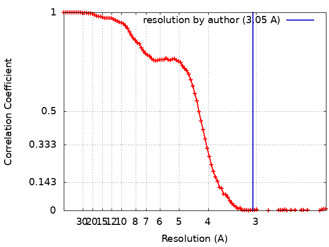
Journal: J Thromb Haemost / Year: 2024 Title: Structural architecture of the acidic region of the B domain of coagulation factor V. Authors: Bassem M Mohammed / Katherine Basore / Brock Summers / Leslie A Pelc / Enrico Di Cera / Abstract: BACKGROUND: Coagulation factor (F)V features an A1-A2-B-A3-C1-C2 domain organization and functions as the inactive precursor of FVa, a component of the prothrombinase complex required for rapid ...BACKGROUND: Coagulation factor (F)V features an A1-A2-B-A3-C1-C2 domain organization and functions as the inactive precursor of FVa, a component of the prothrombinase complex required for rapid thrombin generation in the penultimate step of the coagulation cascade. An intramolecular interaction within the large B domain (residues 710-1545) involves the basic region (BR, residues 963-1008) and acidic region (AR, residues 1493-1537) and locks FV in its inactive state. However, structural information on this important regulatory interaction or on the separate architecture of the AR and BR remains elusive due to conformational disorder of the B domain. OBJECTIVES: To reveal the structure of the BR-AR interaction or of its separate components. METHODS: The structure of FV is solved by cryogenic electron microscopy. RESULTS: A new 3.05 Å resolution cryogenic electron microscopy structure of FV confirms the overall organization of the A and C domains but resolves the segment 1507 to 1545 within a largely ...RESULTS: A new 3.05 Å resolution cryogenic electron microscopy structure of FV confirms the overall organization of the A and C domains but resolves the segment 1507 to 1545 within a largely disordered B domain. The segment contains most of the AR and is organized as recently reported in FV short, a spliced variant of FV with a significantly shorter and less disordered B domain. CONCLUSION: The similar architecture of the AR in FV and FV short provides structural context for physiologically important interactions of this region with the BR in FV and with the basic C-terminal ...CONCLUSION: The similar architecture of the AR in FV and FV short provides structural context for physiologically important interactions of this region with the BR in FV and with the basic C-terminal end of tissue factor pathway inhibitor α in FV short.
pH: 7.4 / Details: 20 mM HEPES, 150 mM NaCl, 5 mM CaCl2
Grid
Model: Quantifoil R2/2 / Material: COPPER / Mesh: 300 / Support film - Material: CARBON / Support film - topology: HOLEY / Pretreatment - Type: PLASMA CLEANING / Pretreatment - Time: 60 sec. / Pretreatment - Atmosphere: OTHER / Pretreatment - Pressure: 0.009300000000000001 kPa
Vitrification
Cryogen name: ETHANE / Chamber humidity: 100 % / Chamber temperature: 277.15 K / Instrument: FEI VITROBOT MARK IV
-
Electron microscopy
Microscope
FEI TITAN
Specialist optics
Energy filter - Name: GIF Bioquantum / Energy filter - Slit width: 20 eV
Image recording
Film or detector model: GATAN K2 SUMMIT (4k x 4k) / Detector mode: COUNTING / Digitization - Dimensions - Width: 4000 pixel / Digitization - Dimensions - Height: 4000 pixel / Number grids imaged: 2 / Number real images: 8429 / Average exposure time: 1.65 sec. / Average electron dose: 66.0 e/Å2 Details: 2 Grids imaged one at 0.1 mg/mL and the second at 0.2 mg/mL using the same acquisition parameters.
Electron beam
Acceleration voltage: 300 kV / Electron source: FIELD EMISSION GUN
In the structure databanks used in Yorodumi, some data are registered as the other names, "COVID-19 virus" and "2019-nCoV". Here are the details of the virus and the list of structure data.
Jan 31, 2019. EMDB accession codes are about to change! (news from PDBe EMDB page)
EMDB accession codes are about to change! (news from PDBe EMDB page)
The allocation of 4 digits for EMDB accession codes will soon come to an end. Whilst these codes will remain in use, new EMDB accession codes will include an additional digit and will expand incrementally as the available range of codes is exhausted. The current 4-digit format prefixed with “EMD-” (i.e. EMD-XXXX) will advance to a 5-digit format (i.e. EMD-XXXXX), and so on. It is currently estimated that the 4-digit codes will be depleted around Spring 2019, at which point the 5-digit format will come into force.
The EM Navigator/Yorodumi systems omit the EMD- prefix.
Related info.:Q: What is EMD? / ID/Accession-code notation in Yorodumi/EM Navigator
Yorodumi is a browser for structure data from EMDB, PDB, SASBDB, etc.
This page is also the successor to EM Navigator detail page, and also detail information page/front-end page for Omokage search.
The word "yorodu" (or yorozu) is an old Japanese word meaning "ten thousand". "mi" (miru) is to see.
Related info.:EMDB / PDB / SASBDB / Comparison of 3 databanks / Yorodumi Search / Aug 31, 2016. New EM Navigator & Yorodumi / Yorodumi Papers / Jmol/JSmol / Function and homology information / Changes in new EM Navigator and Yorodumi
 Movie
Movie Controller

 Yorodumi
 Open data
 Basic information
 Map data
 Sample
 Keywords
 Function and homology information
 Homo sapiens (human)
 Authors
 United States, 5 items
  Citation
 Structure visualization
 Downloads & links
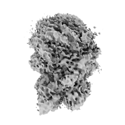 emd_41400.png
 http://ftp.pdbj.org/pub/emdb/structures/EMD-41400


 Z (Sec.)
 Y (Row.)
 X (Col.)


 Sample components
 Processing
 Electron microscopy
 FIELD EMISSION GUN